QUESTION IMAGE
Question
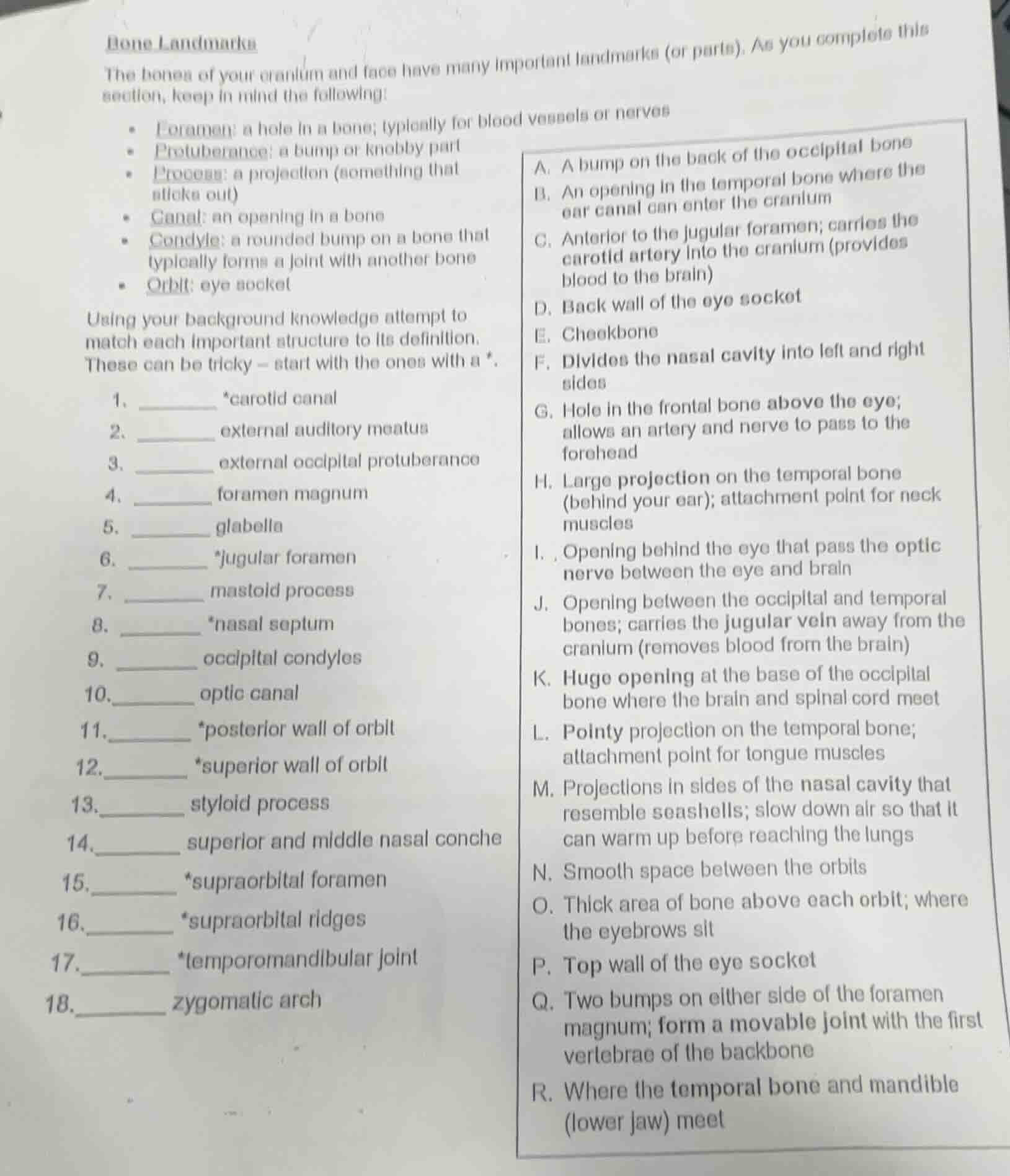
bone landmarks
the bones of your cranium and face have many important landmarks (or parts). as you complete this section, keep in mind the following:
- foramen: a hole in a bone; typically for blood vessels or nerves
- protuberance: a bump or knobby part
- process: a projection (something that sticks out)
- canal: an opening in a bone
- condyle: a rounded bump on a bone that typically forms a joint with another bone
- orbit: eye socket
using your background knowledge attempt to match each important structure to its definition.
these can be tricky – start with the ones with a *.
- _______ *carotid canal
- _______ external auditory meatus
- _______ external occipital protuberance
- _______ foramen magnum
- _______ glabella
- _______ *jugular foramen
- _______ mastoid process
- _______ *nasal septum
- _______ occipital condyles
10._______ optic canal
11._______ *posterior wall of orbit
12._______ *superior wall of orbit
13._______ styloid process
14._______ superior and middle nasal conche
15._______ *supraorbital foramen
16._______ *supraorbital ridges
17._______ *temporomandibular joint
18._______ zygomatic arch
a. a bump on the back of the occipital bone
b. an opening in the temporal bone where the ear canal can enter the cranium
c. anterior to the jugular foramen; carries the carotid artery into the cranium (provides blood to the brain)
d. back wall of the eye socket
e. cheekbone
f. divides the nasal cavity into left and right sides
g. hole in the frontal bone above the eye; allows an artery and nerve to pass to the forehead
h. large projection on the temporal bone (behind your ear); attachment point for neck muscles
i. opening behind the eye that pass the optic nerve between the eye and brain
j. opening between the occipital and temporal bones; carries the jugular vein away from the cranium (removes blood from the brain)
k. huge opening at the base of the occipital bone where the brain and spinal cord meet
l. pointy projection on the temporal bone; attachment point for tongue muscles
m. projections in sides of the nasal cavity that resemble seashells; slow down air so that it can warm up before reaching the lungs
n. smooth space between the orbits
o. thick area of bone above each orbit; where the eyebrows sit
p. top wall of the eye socket
q. two bumps on either side of the foramen magnum; form a movable joint with the first vertebrae of the backbone
r. where the temporal bone and mandible (lower jaw) meet
Each bone structure is matched to its correct definition using anatomical knowledge of cranial and facial bone landmarks:
- The carotid canal is the opening that lets the carotid artery enter the cranium to supply blood to the brain.
- The external auditory meatus is the opening in the temporal bone that connects to the ear canal.
- The external occipital protuberance is a bump on the back of the occipital bone.
- The foramen magnum is the large opening at the base of the occipital bone where the brain connects to the spinal cord.
- The glabella is the smooth space between the two orbits.
- The jugular foramen is the opening between the occipital and temporal bones that carries the jugular vein away from the brain.
- The mastoid process is the large projection on the temporal bone behind the ear, which is an attachment point for neck muscles.
- The nasal septum is the structure that divides the nasal cavity into left and right sides.
- The occipital condyles are the two bumps beside the foramen magnum that form a joint with the first vertebra.
- The optic canal is the opening behind the eye that lets the optic nerve pass between the eye and brain.
- The posterior wall of orbit is the back wall of the eye socket.
- The superior wall of orbit is the top wall of the eye socket.
- The styloid process is the pointy projection on the temporal bone that is an attachment point for tongue muscles.
- The superior and middle nasal conchae are the shell-like projections in the nasal cavity that warm incoming air.
- The supraorbital foramen is the hole in the frontal bone above the eye that allows nerves and arteries to pass to the forehead.
- The supraorbital ridges are the thick bone areas above each orbit where the eyebrows sit.
- The temporomandibular joint is where the temporal bone and lower jaw (mandible) meet.
- The zygomatic arch is the cheekbone.

Snap & solve any problem in the app
Get step-by-step solutions on Sovi AI
Photo-based solutions with guided steps
Explore more problems and detailed explanations
- C. Anterior to the jugular foramen; carries the carotid artery into the cranium (provides blood to the brain)
- B. An opening in the temporal bone where the ear canal can enter the cranium
- A. A bump on the back of the occipital bone
- K. Huge opening at the base of the occipital bone where the brain and spinal cord meet
- N. Smooth space between the orbits
- J. Opening between the occipital and temporal bones; carries the jugular vein away from the cranium (removes blood from the brain)
- H. Large projection on the temporal bone (behind your ear); attachment point for neck muscles
- F. Divides the nasal cavity into left and right sides
- Q. Two bumps on either side of the foramen magnum; form a movable joint with the first vertebrae of the backbone
- I. Opening behind the eye that pass the optic nerve between the eye and brain
- D. Back wall of the eye socket
- P. Top wall of the eye socket
- L. Pointy projection on the temporal bone; attachment point for tongue muscles
- M. Projections in sides of the nasal cavity that resemble seashells; slow down air so that it can warm up before reaching the lungs
- G. Hole in the frontal bone above the eye; allows an artery and nerve to pass to the forehead
- O. Thick area of bone above each orbit; where the eyebrows sit
- R. Where the temporal bone and mandible (lower jaw) meet
- E. Cheekbone