QUESTION IMAGE
Question
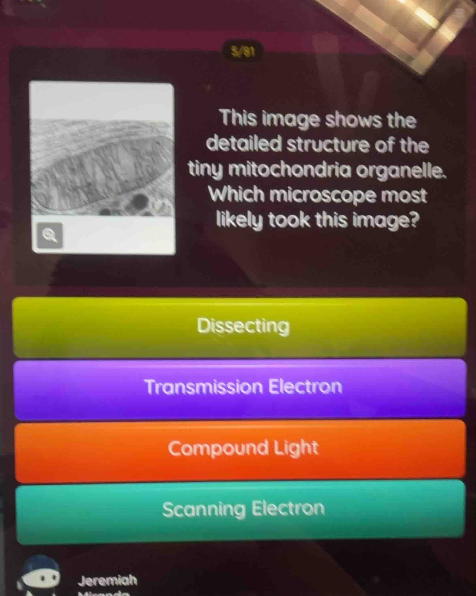
this image shows the detailed structure of the tiny mitochondria organelle. which microscope most likely took this image? dissecting transmission electron compound light scanning electron
Brief Explanations
To determine the microscope that captured the detailed structure of mitochondria, we analyze each option:
- Dissecting microscopes have low magnification and are used for viewing larger, opaque objects (e.g., insects, plant parts), not small organelles like mitochondria. Eliminate this option.
- Transmission Electron Microscopes (TEMs) use electrons to pass through thin specimens, allowing for high - resolution imaging of internal structures of cells and organelles (like mitochondria). This matches the need for detailed internal structure visualization.
- Compound light microscopes use visible light and have limited magnification (up to ~1000x), which is insufficient to show the detailed internal structure of mitochondria. Eliminate this option.
- Scanning Electron Microscopes (SEMs) are used to view the surface structure of specimens, not the internal structure of organelles. Eliminate this option.

Snap & solve any problem in the app
Get step-by-step solutions on Sovi AI
Photo-based solutions with guided steps
Explore more problems and detailed explanations
B. Transmission Electron