QUESTION IMAGE
Question
- nucleus
- nucleolus
- cytoplasm
- cell membranes
- lysosome
- endoplasmic reticulum
- golgi apparatus
- mitochondria
- ribosome
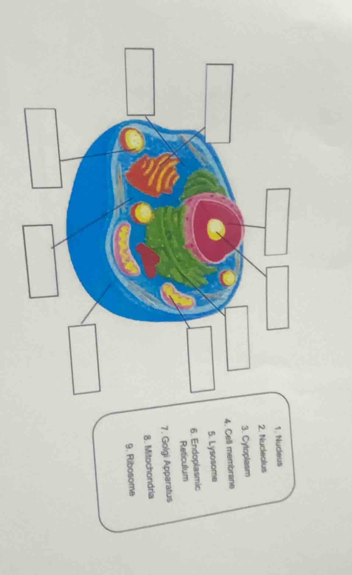
To solve this cell structure labeling problem, we identify each organelle by its typical appearance:
Step 1: Identify the Nucleus (1)
The large, pinkish - red structure with a yellow center (nucleolus) is the nucleus. So the label for the nucleus - like structure is 1. Nucleus.
Step 2: Identify the Nucleolus (2)
The small yellow dot inside the nucleus is the nucleolus. So the label for this small yellow dot is 2. Nucleolus.
Step 3: Identify the Cytoplasm (3)
The blue - colored region filling the space between the cell membrane and the organelles is the cytoplasm. So the label for this blue region is 3. Cytoplasm.
Step 4: Identify the Cell Membrane (4)
The outer boundary of the cell (the blue outer layer) is the cell membrane. So the label for this outer layer is 4. Cell Membrane.
Step 5: Identify the Lysosome (5)
The small, round, yellow - colored structures (similar to small vesicles) are lysosomes. So the label for these small round structures is 5. Lysosome.
Step 6: Identify the Endoplasmic Reticulum (6)
The network - like, membrane - bound structures (can be rough or smooth, here the network - like ones) are the endoplasmic reticulum. So the label for these network - like structures is 6. Endoplasmic Reticulum.
Step 7: Identify the Golgi Apparatus (7)
The stack - like, red - orange colored structure is the Golgi apparatus. So the label for this stack - like structure is 7. Golgi Apparatus.
Step 8: Identify the Mitochondria (8)
The oval - shaped, pink - yellow colored structures with inner folds are mitochondria. So the label for these oval - shaped structures is 8. Mitochondria.
Step 9: Identify the Ribosome (9)
The tiny dots (if visible, or in the context of the cell, the small structures associated with the endoplasmic reticulum or free in cytoplasm) are ribosomes. So the label for these tiny dots is 9. Ribosome.
(Note: The exact placement of each label depends on the position of the boxes in the diagram. For example, if a box is pointing to the nucleus, it gets label 1; if pointing to the nucleolus, label 2, and so on following the above identifications.)

Snap & solve any problem in the app
Get step-by-step solutions on Sovi AI
Photo-based solutions with guided steps
Explore more problems and detailed explanations
To solve this cell structure labeling problem, we identify each organelle by its typical appearance:
Step 1: Identify the Nucleus (1)
The large, pinkish - red structure with a yellow center (nucleolus) is the nucleus. So the label for the nucleus - like structure is 1. Nucleus.
Step 2: Identify the Nucleolus (2)
The small yellow dot inside the nucleus is the nucleolus. So the label for this small yellow dot is 2. Nucleolus.
Step 3: Identify the Cytoplasm (3)
The blue - colored region filling the space between the cell membrane and the organelles is the cytoplasm. So the label for this blue region is 3. Cytoplasm.
Step 4: Identify the Cell Membrane (4)
The outer boundary of the cell (the blue outer layer) is the cell membrane. So the label for this outer layer is 4. Cell Membrane.
Step 5: Identify the Lysosome (5)
The small, round, yellow - colored structures (similar to small vesicles) are lysosomes. So the label for these small round structures is 5. Lysosome.
Step 6: Identify the Endoplasmic Reticulum (6)
The network - like, membrane - bound structures (can be rough or smooth, here the network - like ones) are the endoplasmic reticulum. So the label for these network - like structures is 6. Endoplasmic Reticulum.
Step 7: Identify the Golgi Apparatus (7)
The stack - like, red - orange colored structure is the Golgi apparatus. So the label for this stack - like structure is 7. Golgi Apparatus.
Step 8: Identify the Mitochondria (8)
The oval - shaped, pink - yellow colored structures with inner folds are mitochondria. So the label for these oval - shaped structures is 8. Mitochondria.
Step 9: Identify the Ribosome (9)
The tiny dots (if visible, or in the context of the cell, the small structures associated with the endoplasmic reticulum or free in cytoplasm) are ribosomes. So the label for these tiny dots is 9. Ribosome.
(Note: The exact placement of each label depends on the position of the boxes in the diagram. For example, if a box is pointing to the nucleus, it gets label 1; if pointing to the nucleolus, label 2, and so on following the above identifications.)