QUESTION IMAGE
Question
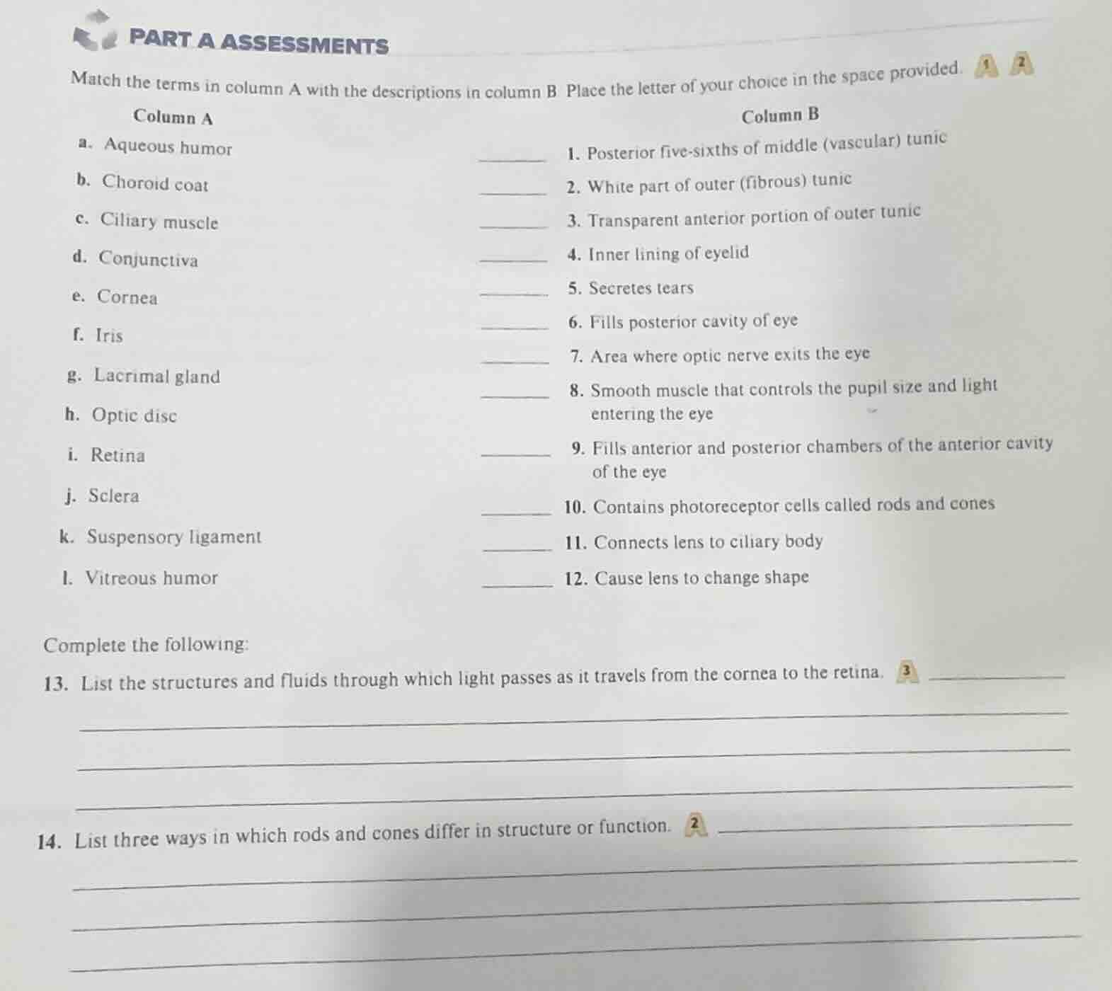
part a assessments
match the terms in column a with the descriptions in column b place the letter of your choice in the space provided.
column a
a. aqueous humor
b. choroid coat
c. ciliary muscle
d. conjunctiva
e. cornea
f. iris
g. lacrimal gland
h. optic disc
i. retina
j. sclera
k. suspensory ligament
l. vitreous humor
column b
- posterior five - sixths of middle (vascular) tunic
- white part of outer (fibrous) tunic
- transparent anterior portion of outer tunic
- inner lining of eyelid
- secretes tears
- fills posterior cavity of eye
- area where optic nerve exits the eye
- smooth muscle that controls the pupil size and light entering the eye
- fills anterior and posterior chambers of the anterior cavity of the eye
- contains photoreceptor cells called rods and cones
- connects lens to ciliary body
- cause lens to change shape
complete the following:
- list the structures and fluids through which light passes as it travels from the cornea to the retina.
- list three ways in which rods and cones differ in structure or function.
Matching Section:
Each term from Column A is paired with its corresponding anatomical description in Column B based on human eye anatomy.
Question 13:
Light travels through a sequence of transparent eye structures/fluids from the cornea to the retina, following the eye's optical pathway.
Question 14:
Rods and cones differ in multiple structural and functional aspects related to vision; three key distinct traits are selected.

Snap & solve any problem in the app
Get step-by-step solutions on Sovi AI
Photo-based solutions with guided steps
Explore more problems and detailed explanations
Matching Answers:
- b. Choroid coat
- j. Sclera
- e. Cornea
- d. Conjunctiva
- g. Lacrimal gland
- l. Vitreous humor
- h. Optic disc
- f. Iris
- a. Aqueous humor
- i. Retina
- k. Suspensory ligament
- c. Ciliary muscle
Question 13:
- Aqueous humor
- Pupil (opening in the iris)
- Lens
- Vitreous humor
Question 14:
- Function: Rods detect dim light (night vision); cones detect bright light and color.
- Distribution: Rods are more dense in the peripheral retina; cones are concentrated in the fovea centralis.
- Structure: Rods have a long, thin outer segment; cones have a shorter, tapering outer segment.