QUESTION IMAGE
Question
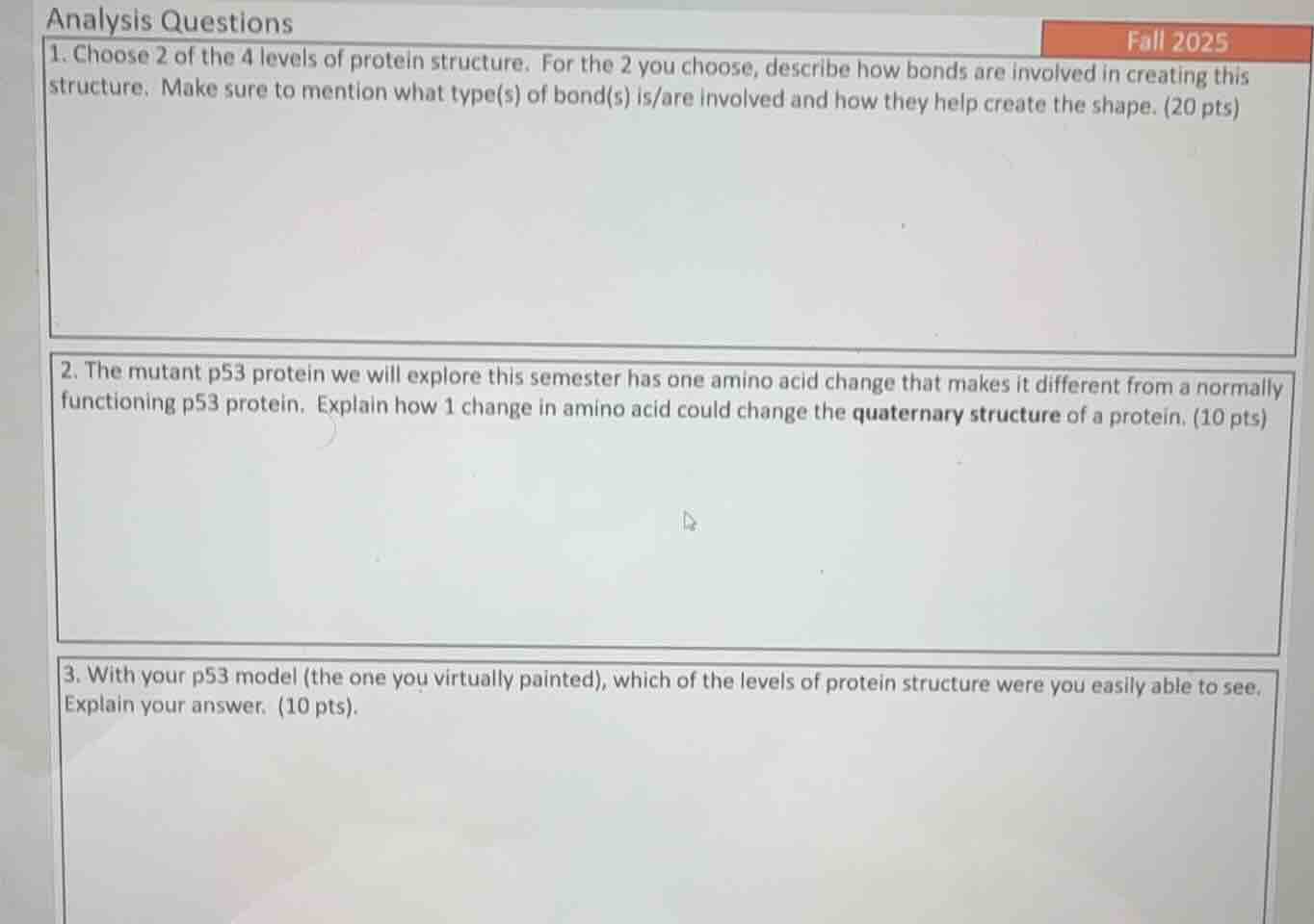
analysis questions
- choose 2 of the 4 levels of protein structure. for the 2 you choose, describe how bonds are involved in creating this structure. make sure to mention what type(s) of bond(s) is/are involved and how they help create the shape. (20 pts)
- the mutant p53 protein we will explore this semester has one amino acid change that makes it different from a normally functioning p53 protein. explain how 1 change in amino acid could change the quaternary structure of a protein. (10 pts)
- with your p53 model (the one you virtually painted), which of the levels of protein structure were you easily able to see. explain your answer. (10 pts).
Brief Explanations
- For the first question, two protein structure levels are selected, with bond roles explained:
- Primary structure: Relies on peptide bonds linking amino acids in a linear chain, forming the core sequence.
- Tertiary structure: Uses hydrogen bonds, disulfide bridges, hydrophobic interactions, and ionic bonds to fold the secondary structure into a 3D shape.
- A single amino acid change can alter the surface chemistry (like charge or hydrophobicity) of a protein subunit. This disrupts the non-covalent bonds (hydrogen bonds, hydrophobic interactions, ionic bonds) that hold multiple subunits together in the quaternary structure, preventing proper subunit assembly or causing incorrect association.
- On a virtual p53 model, the tertiary and quaternary structures are easiest to see. The tertiary structure is visible as the distinct 3D folded shape of individual p53 subunits, while the quaternary structure is observable as the arrangement of multiple p53 subunits bonded together to form the full protein complex.

Snap & solve any problem in the app
Get step-by-step solutions on Sovi AI
Photo-based solutions with guided steps
Explore more problems and detailed explanations
1.
- Primary Protein Structure: Peptide bonds form between the carboxyl group of one amino acid and the amino group of the next, creating a linear amino acid chain. This covalent bond establishes the core sequence that defines the protein's basic identity.
- Tertiary Protein Structure: Hydrogen bonds form between polar R-groups, disulfide bridges form between cysteine residues, hydrophobic interactions cluster nonpolar R-groups inward, and ionic bonds form between charged R-groups. These bonds work together to fold the secondary structure into a stable, unique 3D shape.
- A single amino acid substitution can alter the chemical properties (e.g., charge, hydrophobicity, size) of the protein's surface. This disrupts the specific non-covalent interactions (hydrogen bonds, hydrophobic forces, ionic bonds) that normally mediate the binding of protein subunits to one another. As a result, subunits may no longer recognize or associate with each other correctly, leading to a defective or altered quaternary structure.
- The tertiary and quaternary levels of protein structure are easily visible on the virtual p53 model:
- Tertiary structure: Visible as the distinct, folded 3D shape of each individual p53 subunit, with recognizable loops, helices, and folded regions.
- Quaternary structure: Visible as the assembled complex of multiple p53 subunits bonded together, showing how the individual 3D subunits interact to form the full functional protein.