QUESTION IMAGE
Question
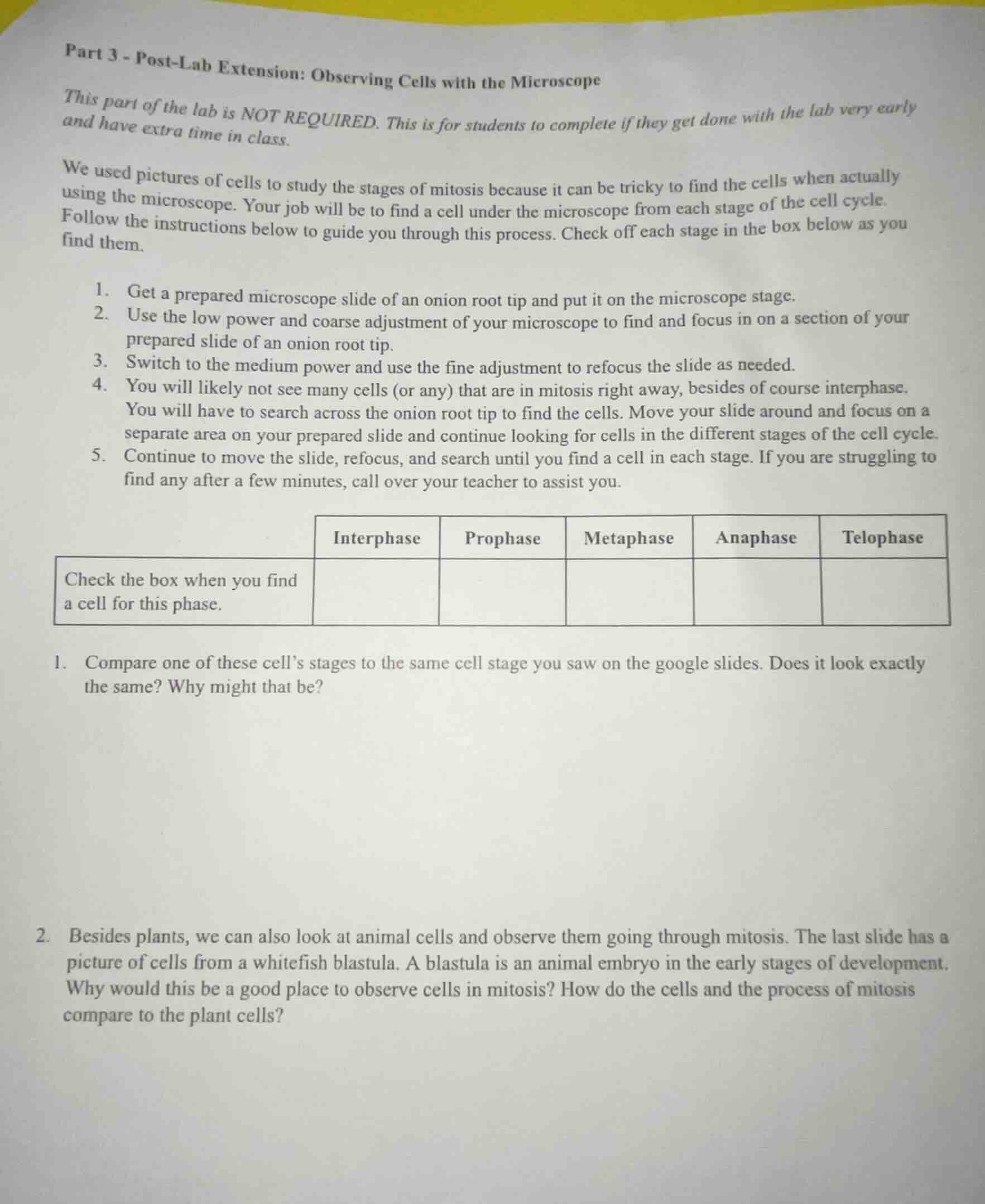
part 3 - post-lab extension: observing cells with the microscope
this part of the lab is not required. this is for students to complete if they get done with the lab very early and have extra time in class.
we used pictures of cells to study the stages of mitosis because it can be tricky to find the cells when actually using the microscope. your job will be to find a cell under the microscope from each stage of the cell cycle. follow the instructions below to guide you through this process. check off each stage in the box below as you find them.
- get a prepared microscope slide of an onion root tip and put it on the microscope stage.
- use the low power and coarse adjustment of your microscope to find and focus in on a section of your prepared slide of an onion root tip.
- switch to the medium power and use the fine adjustment to refocus the slide as needed.
- you will likely not see many cells (or any) that are in mitosis right away, besides of course interphase. you will have to search across the onion root tip to find the cells. move your slide around and focus on a separate area on your prepared slide and continue looking for cells in the different stages of the cell cycle.
- continue to move the slide, refocus, and search until you find a cell in each stage. if you are struggling to find any after a few minutes, call over your teacher to assist you.
| check the box when you find a cell for this phase. | interphase | prophase | metaphase | anaphase | telophase |
|---|
- compare one of these cell’s stages to the same cell stage you saw on the google slides. does it look exactly the same? why might that be?
- besides plants, we can also look at animal cells and observe them going through mitosis. the last slide has a picture of cells from a whitefish blastula. a blastula is an animal embryo in the early stages of development. why would this be a good place to observe cells in mitosis? how do the cells and the process of mitosis compare to the plant cells?
Brief Explanations
- For the first question: The observed cell stage will not look exactly like the Google Slides image. Prepared real slides have natural cell variation, minor slide preparation artifacts, and microscope resolution limits, while Google Slides images are often simplified, idealized illustrations of the stages.
- For the second question: A whitefish blastula is good for observing mitosis because it is an early embryo with rapid, continuous cell division to support growth, so many cells are in mitotic stages. Compared to plant cells: whitefish (animal) cells lack a cell wall, so they round up during mitosis; they use a cleavage furrow to split cytoplasm during cytokinesis, while plant cells form a cell plate; the overall mitotic steps (prophase, metaphase, anaphase, telophase) are the same, but the structural differences of the cells create visible distinctions in the process.

Snap & solve any problem in the app
Get step-by-step solutions on Sovi AI
Photo-based solutions with guided steps
Explore more problems and detailed explanations
- No, it will not look exactly the same. Real cells have natural variation, slide artifacts, and microscope limits, while the Google Slides images are idealized illustrations.
- A whitefish blastula is a good sample because its embryonic cells divide rapidly and continuously, so many cells are in mitosis. Compared to plant cells: animal cells lack a cell wall, round up during mitosis, and use a cleavage furrow for cytokinesis, while plant cells form a cell plate; the core mitotic stages are identical, but cellular structural differences create visible process distinctions.