QUESTION IMAGE
Question
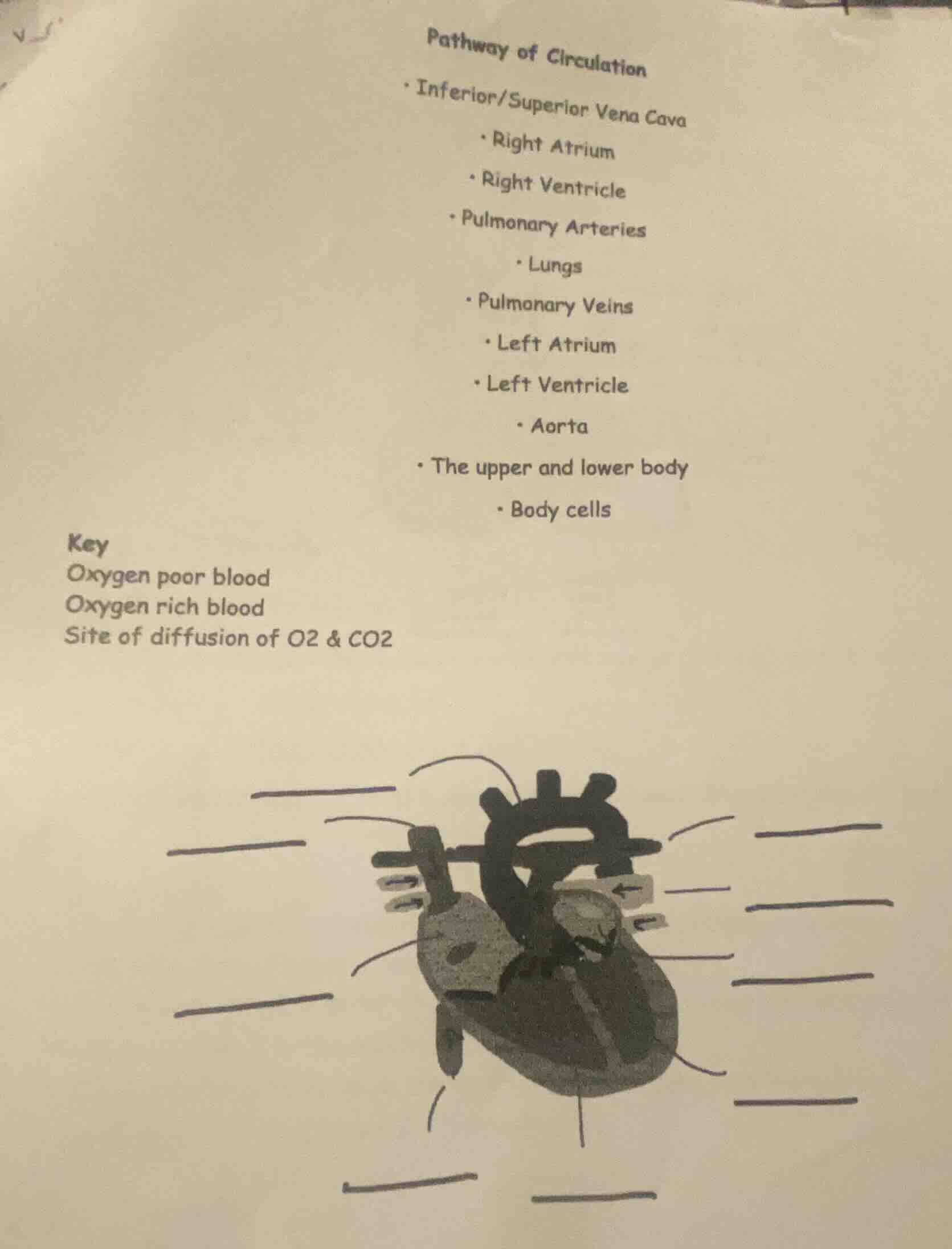
pathway of circulation
- inferior/superior vena cava
- right atrium
- right ventricle
- pulmonary arteries
- lungs
- pulmonary veins
- left atrium
- left ventricle
- aorta
- the upper and lower body
- body cells
key
oxygen poor blood
oxygen rich blood
site of diffusion of o2 & co2
Since the problem (presumably labeling the diagram of the circulatory system) is about the human circulatory system, the relevant subfield is Biology (under Natural Science). Here's a guide to labeling the diagram based on the "Pathway of Circulation" provided:
Step 1: Identify the structures from the pathway
- The large vessels bringing deoxygenated blood to the heart: Inferior/Superior Vena Cava.
- The right upper chamber of the heart: Right Atrium.
- The right lower chamber of the heart: Right Ventricle.
- Vessels carrying deoxygenated blood to the lungs: Pulmonary Arteries.
- Organs where gas exchange occurs: Lungs (site of diffusion of O₂ & CO₂).
- Vessels carrying oxygenated blood from the lungs: Pulmonary Veins.
- The left upper chamber of the heart: Left Atrium.
- The left lower chamber of the heart: Left Ventricle.
- The large vessel carrying oxygenated blood to the body: Aorta.
- The destination of the aorta's blood: The upper and lower body (leading to Body cells, where gas exchange also occurs at the tissue level).
Step 2: Match to the diagram
Assuming the diagram has lines pointing to each structure:
- The vessels entering the top-right of the heart (right atrium area) are the Inferior/Superior Vena Cava (carrying oxygen - poor blood).
- The chamber they empty into is the Right Atrium.
- From the right atrium, blood moves to the Right Ventricle.
- From the right ventricle, the vessel leading out (towards the lungs) is the Pulmonary Arteries (oxygen - poor blood).
- The structure the pulmonary arteries lead to is the Lungs (site of O₂ and CO₂ diffusion, oxygen - poor blood becomes oxygen - rich here).
- The vessels leading from the lungs back to the heart (left atrium area) are the Pulmonary Veins (oxygen - rich blood).
- The chamber they empty into is the Left Atrium.
- From the left atrium, blood moves to the Left Ventricle.
- From the left ventricle, the large vessel leading out (to the body) is the Aorta (oxygen - rich blood).
- The aorta branches to supply the The upper and lower body, and finally the blood reaches Body cells (where oxygen is delivered and carbon dioxide is picked up, oxygen - rich blood becomes oxygen - poor).
If you were to label each line in the diagram:
- If a line points to the large vessels entering the right side of the heart: Label as "Inferior/Superior Vena Cava (Oxygen poor blood)".
- If a line points to the right upper heart chamber: Label as "Right Atrium".
- If a line points to the right lower heart chamber: Label as "Right Ventricle".
- If a line points to the vessels going from the right ventricle to the lungs: Label as "Pulmonary Arteries (Oxygen poor blood)".
- If a line points to the lung structures: Label as "Lungs (Site of diffusion of O₂ & CO₂)".
- If a line points to the vessels going from the lungs to the left side of the heart: Label as "Pulmonary Veins (Oxygen rich blood)".
- If a line points to the left upper heart chamber: Label as "Left Atrium".
- If a line points to the left lower heart chamber: Label as "Left Ventricle".
- If a line points to the large vessel going from the left ventricle to the body: Label as "Aorta (Oxygen rich blood)".
- If a line points to the areas representing the body (or the small vessels in the body region): Label as "The upper and lower body (leads to Body cells, site of tissue - level diffusion)".
- If a line points to the small structures representing cells: Label as "Body cells (Site of diffusion of O₂ & CO₂ at tissue level)".

Snap & solve any problem in the app
Get step-by-step solutions on Sovi AI
Photo-based solutions with guided steps
Explore more problems and detailed explanations
Since the problem (presumably labeling the diagram of the circulatory system) is about the human circulatory system, the relevant subfield is Biology (under Natural Science). Here's a guide to labeling the diagram based on the "Pathway of Circulation" provided:
Step 1: Identify the structures from the pathway
- The large vessels bringing deoxygenated blood to the heart: Inferior/Superior Vena Cava.
- The right upper chamber of the heart: Right Atrium.
- The right lower chamber of the heart: Right Ventricle.
- Vessels carrying deoxygenated blood to the lungs: Pulmonary Arteries.
- Organs where gas exchange occurs: Lungs (site of diffusion of O₂ & CO₂).
- Vessels carrying oxygenated blood from the lungs: Pulmonary Veins.
- The left upper chamber of the heart: Left Atrium.
- The left lower chamber of the heart: Left Ventricle.
- The large vessel carrying oxygenated blood to the body: Aorta.
- The destination of the aorta's blood: The upper and lower body (leading to Body cells, where gas exchange also occurs at the tissue level).
Step 2: Match to the diagram
Assuming the diagram has lines pointing to each structure:
- The vessels entering the top-right of the heart (right atrium area) are the Inferior/Superior Vena Cava (carrying oxygen - poor blood).
- The chamber they empty into is the Right Atrium.
- From the right atrium, blood moves to the Right Ventricle.
- From the right ventricle, the vessel leading out (towards the lungs) is the Pulmonary Arteries (oxygen - poor blood).
- The structure the pulmonary arteries lead to is the Lungs (site of O₂ and CO₂ diffusion, oxygen - poor blood becomes oxygen - rich here).
- The vessels leading from the lungs back to the heart (left atrium area) are the Pulmonary Veins (oxygen - rich blood).
- The chamber they empty into is the Left Atrium.
- From the left atrium, blood moves to the Left Ventricle.
- From the left ventricle, the large vessel leading out (to the body) is the Aorta (oxygen - rich blood).
- The aorta branches to supply the The upper and lower body, and finally the blood reaches Body cells (where oxygen is delivered and carbon dioxide is picked up, oxygen - rich blood becomes oxygen - poor).
If you were to label each line in the diagram:
- If a line points to the large vessels entering the right side of the heart: Label as "Inferior/Superior Vena Cava (Oxygen poor blood)".
- If a line points to the right upper heart chamber: Label as "Right Atrium".
- If a line points to the right lower heart chamber: Label as "Right Ventricle".
- If a line points to the vessels going from the right ventricle to the lungs: Label as "Pulmonary Arteries (Oxygen poor blood)".
- If a line points to the lung structures: Label as "Lungs (Site of diffusion of O₂ & CO₂)".
- If a line points to the vessels going from the lungs to the left side of the heart: Label as "Pulmonary Veins (Oxygen rich blood)".
- If a line points to the left upper heart chamber: Label as "Left Atrium".
- If a line points to the left lower heart chamber: Label as "Left Ventricle".
- If a line points to the large vessel going from the left ventricle to the body: Label as "Aorta (Oxygen rich blood)".
- If a line points to the areas representing the body (or the small vessels in the body region): Label as "The upper and lower body (leads to Body cells, site of tissue - level diffusion)".
- If a line points to the small structures representing cells: Label as "Body cells (Site of diffusion of O₂ & CO₂ at tissue level)".