QUESTION IMAGE
Question
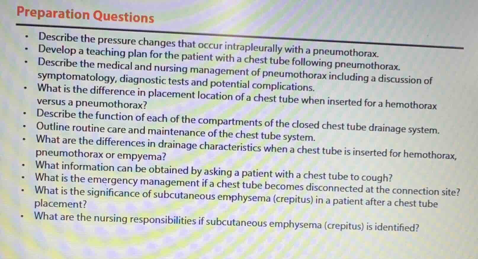
preparation questions
- describe the pressure changes that occur intrapleurally with a pneumothorax.
- develop a teaching plan for the patient with a chest tube following pneumothorax.
- describe the medical and nursing management of pneumothorax including a discussion of symptomatology, diagnostic tests and potential complications.
- what is the difference in placement location of a chest tube when inserted for a hemothorax versus a pneumothorax?
- describe the function of each of the compartments of the closed chest tube drainage system.
- outline routine care and maintenance of the chest tube system.
- what are the differences in drainage characteristics when a chest tube is inserted for hemothorax, pneumothorax or empyema?
- what information can be obtained by asking a patient with a chest tube to cough?
- what is the emergency management if a chest tube becomes disconnected at the connection site?
- what is the significance of subcutaneous emphysema (crepitus) in a patient after a chest tube placement?
- what are the nursing responsibilities if subcutaneous emphysema (crepitus) is identified?
Brief Explanations
- Intrapleural pressure changes with pneumothorax: Normally, intrapleural pressure is negative relative to atmospheric pressure (-4 to -6 cm H₂O at rest). A pneumothorax lets air enter the pleural space, equalizing pressure to atmospheric pressure (0 cm H₂O), collapsing the lung.
- Chest tube patient teaching plan: Covers purpose of the tube, activity restrictions, signs of complications (pain, crepitus, air leak changes), deep breathing/coughing techniques, and care of the drainage system.
- Pneumothorax medical/nursing management:
- Medical: Supplemental O₂, needle decompression for tension pneumothorax, chest tube insertion for evacuation.
- Nursing: Monitor vital signs, respiratory status, chest tube drainage, assess for complications, assist with mobility, provide education.
- Symptomatology: Dyspnea, chest pain, tachypnea, decreased breath sounds.
- Diagnostic tests: Chest X-ray, CT scan, pulse oximetry.
- Complications: Tension pneumothorax, hemothorax, infection, subcutaneous emphysema.
- Chest tube placement: hemothorax vs pneumothorax:
- Pneumothorax: Inserted in the 2nd intercostal space, midclavicular line (to evacuate air).
- Hemothorax: Inserted in the 4th-5th intercostal space, midaxillary line (to drain fluid/blood, which settles inferiorly).
- Closed chest tube drainage system compartments:
- Collection chamber: Catches drained air/fluid, measures volume.
- Water-seal chamber: Prevents backflow of air into the pleural space; fluctuates with respiration (tidaling) and shows air leaks (bubbling).
- Suction control chamber: Regulates the amount of negative pressure applied to the pleural space (either water-based or dry suction).
- Chest tube routine care: Monitor drainage (color, amount, consistency), check for air leaks, keep the system below chest level, maintain patency (avoid kinks/clogs), assess insertion site for infection/crepitus, monitor respiratory status, and ensure suction is set correctly.
- Drainage characteristics by condition:
- Hemothorax: Bloody, serosanguineous drainage, initially large volume that decreases over time.
- Pneumothorax: Primarily air drainage (visible bubbling in water-seal, minimal fluid).
- Empyema: Thick, cloudy, purulent, foul-smelling fluid (infected pleural effusion).
- Information from having a patient cough: Assesses patency of the chest tube (drainage increases with cough), checks for air leaks (increased bubbling), evaluates lung re-expansion, and helps mobilize secretions.
- Chest tube disconnection emergency management: Immediately clamp the tube close to the insertion site, clean the disconnected ends with sterile solution, reconnect the tube, or if unable to reconnect, place the end of the tube in a container of sterile water to create a temporary water seal, then notify the provider.
- Significance of subcutaneous emphysema post chest tube placement: Indicates air has leaked from the pleural space into the subcutaneous tissue, often from a loose insertion site, air leak in the chest tube system, or incomplete lung re-expansion. It can be mild (localized) or severe (spread to face/neck, compromising airway).
- Nursing responsibilities for subcutaneous emphysema: Monitor the extent of crepitus (mark borders to track changes), assess respiratory status, check chest tube system for leaks, ensure the tube is patent, notify the provider, and document findings.

Snap & solve any problem in the app
Get step-by-step solutions on Sovi AI
Photo-based solutions with guided steps
Explore more problems and detailed explanations
- Intrapleural pressure shifts from negative (-4 to -6 cm H₂O) to atmospheric (0 cm H₂O), collapsing the affected lung.
- Teaching plan includes: purpose of chest tube, activity limits, complication recognition, breathing exercises, and drainage system care.
- Medical: O₂ therapy, needle decompression (tension pneumo), chest tube insertion. Nursing: monitor vitals/respiratory status, assess drainage, prevent complications, patient education. Symptoms: dyspnea, chest pain, decreased breath sounds. Diagnostics: chest X-ray, pulse oximetry. Complications: tension pneumothorax, infection, hemothorax.
- Pneumothorax: 2nd intercostal space, midclavicular line. Hemothorax: 4th-5th intercostal space, midaxillary line.
- Collection chamber: collects/measures drained fluid/air; Water-seal chamber: prevents air backflow, detects leaks; Suction control chamber: regulates negative pressure.
- Monitor drainage, check for leaks, keep system below chest, avoid kinks, assess insertion site, verify suction settings, monitor respiratory status.
- Hemothorax: bloody, serosanguineous, large initial volume; Pneumothorax: mostly air, minimal fluid, bubbling; Empyema: thick, purulent, foul-smelling fluid.
- Evaluates chest tube patency, detects air leaks, assesses lung re-expansion, mobilizes secretions.
- Clamp tube near insertion site, clean ends, reconnect or place in sterile water seal, notify provider.
- Signals air leakage from pleural space into subcutaneous tissue; may indicate loose site, tube leak, or incomplete lung re-expansion.
- Track crepitus borders, assess respiratory status, check chest tube for leaks, notify provider, document findings.