QUESTION IMAGE
Question
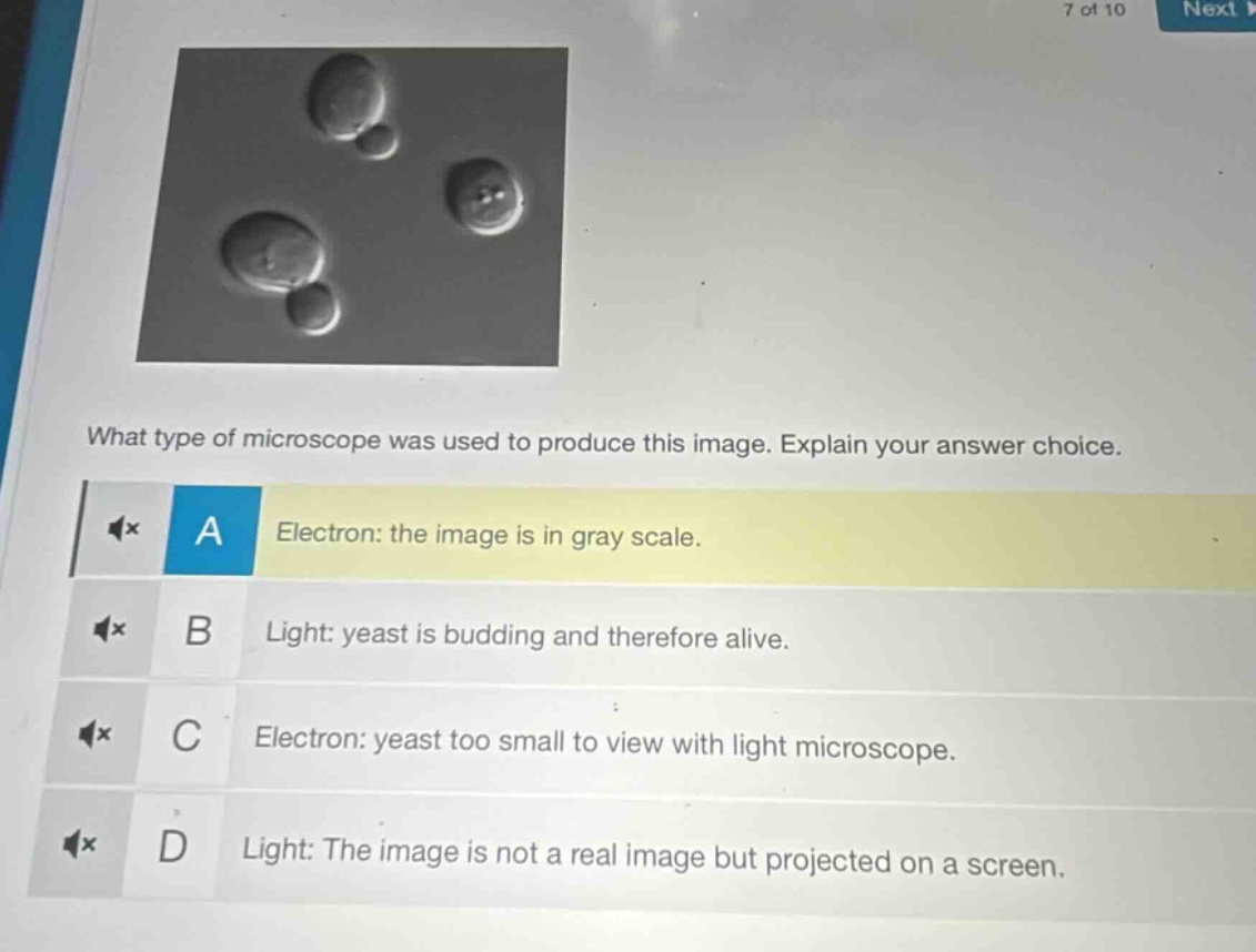
what type of microscope was used to produce this image. explain your answer choice. a electron: the image is in gray scale. b light: yeast is budding and therefore alive. c electron: yeast too small to view with light microscope. d light: the image is not a real image but projected on a screen.
Brief Explanations
To determine the microscope type, we analyze each option:
- Option A: Electron microscopes kill specimens (can't view live budding), and grayscale isn't unique to electron (light can too). Eliminate A.
- Option B: Light microscopes can view live specimens. Yeast budding shows it's alive, which is possible with light (electron can't view live). This fits.
- Option C: Yeast is visible with light microscopes (size ~5 - 10 μm, light can resolve). Eliminate C.
- Option D: The "projected on screen" reasoning is incorrect (both can project, and live viewing is key here). Eliminate D.

Snap & solve any problem in the app
Get step-by-step solutions on Sovi AI
Photo-based solutions with guided steps
Explore more problems and detailed explanations
B. Light: yeast is budding and therefore alive.