QUESTION IMAGE
Question
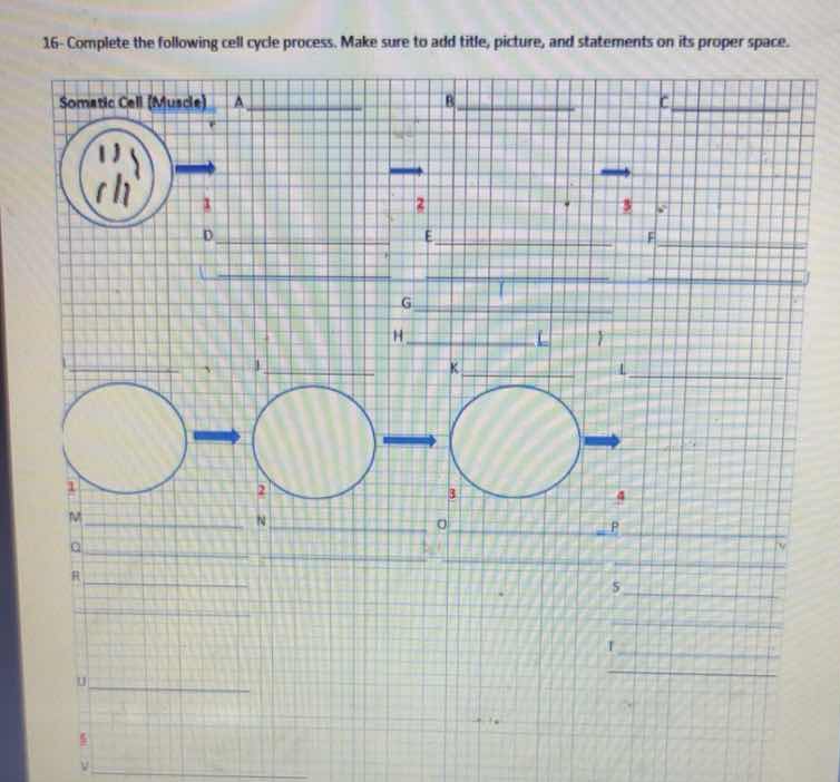
16- complete the following cell cycle process. make sure to add title, picture, and statements on its proper space.
somatic cell (muscle)
Brief Explanations
This task involves mapping the stages of the somatic (muscle) cell cycle, including interphase and mitotic stages, with corresponding labels for each phase, key events, and cell illustrations.
- The top row tracks nuclear/chromosome changes, while the bottom row tracks cell growth and division.
- Each labeled section corresponds to a phase of the cell cycle, its key characteristics, and visual representation.

Snap & solve any problem in the app
Get step-by-step solutions on Sovi AI
Photo-based solutions with guided steps
Explore more problems and detailed explanations
Top Section (Chromosome/Nuclear Cycle)
Title: Chromosomal Changes in Muscle Cell Cycle
- A: Interphase (G1 + S + G2)
- D: Cell grows, DNA replicates
- Picture: Cell with intact nucleus, uncondensed chromosomes (as shown in the first circle)
- B: Prophase
- E: Chromosomes condense, nucleus breaks down
- Picture: Draw condensed chromosomes scattered in the cell, no nuclear envelope
- C: Metaphase
- F: Chromosomes line up at cell equator
- Picture: Draw condensed chromosomes aligned in the center of the cell
- G: Anaphase
- H: Sister chromatids separate, move to poles
- Picture: Draw chromatids pulled apart toward opposite ends of the cell
- K: Telophase
- L: Nuclei reform, chromosomes decondense
- Picture: Draw two new nuclei forming at opposite ends, chromosomes uncoiling
Bottom Section (Cell Growth & Division)
Title: Cytoplasmic Changes in Muscle Cell Cycle
- M: G1 Phase
- Q: Cell grows, performs normal functions
- Picture: Small, single cell (first circle)
- N: S + G2 Phase
- R: DNA replicates, cell prepares to divide
- Picture: Larger single cell (second circle)
- O: Mitosis (Prophase - Telophase)
- T: Nucleus divides, chromosomes separate
- Picture: Large cell with dividing nucleus (third circle)
- P: Cytokinesis
- U: Cytoplasm splits, two new cells form
- Picture: Two identical small muscle cells (final stage)