QUESTION IMAGE
Question
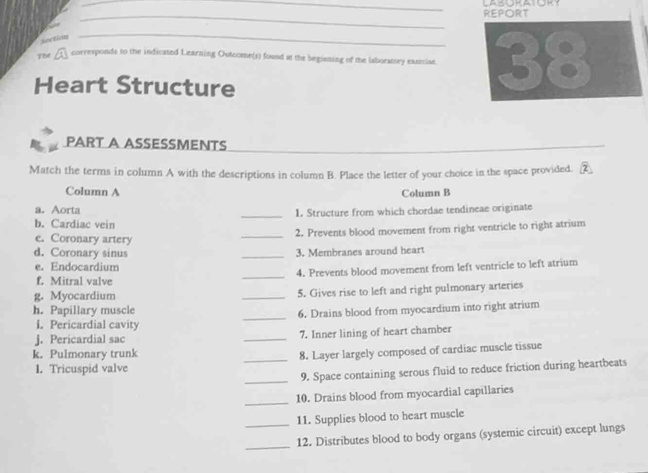
laboratory report
section
the corresponds to the indicated learning outcome(s) found at the beginning of the laboratory exercise.
38
heart structure
part a assessments
match the terms in column a with the descriptions in column b. place the letter of your choice in the space provided.
column a
column b
a. aorta
- structure from which chordae tendineae originate
b. cardiac vein
- prevents blood movement from right ventricle to right atrium
c. coronary artery
- membranes around heart
d. coronary sinus
- prevents blood movement from left ventricle to left atrium
e. endocardium
- gives rise to left and right pulmonary arteries
f. mitral valve
- drains blood from myocardium into right atrium
g. myocardium
- inner lining of heart chamber
h. papillary muscle
- layer largely composed of cardiac muscle tissue
i. pericardial cavity
- space containing serous fluid to reduce friction during heartbeats
j. pericardial sac
- drains blood from myocardial capillaries
k. pulmonary trunk
- supplies blood to heart muscle
l. tricuspid valve
- distributes blood to body organs (systemic circuit) except lungs
Each term in Column A is matched to its corresponding anatomical function/description in Column B based on human heart structure and physiology:
- Papillary muscles anchor chordae tendineae.
- Tricuspid valve controls right atrioventricular flow.
- Pericardial sac encases the heart.
- Mitral valve controls left atrioventricular flow.
- Pulmonary trunk splits into pulmonary arteries.
- Coronary sinus drains myocardium to right atrium.
- Endocardium lines heart chambers.
- Myocardium is cardiac muscle tissue.
- Pericardial cavity holds serous fluid for friction reduction.
- Cardiac veins drain myocardial capillaries.
- Coronary arteries supply blood to heart muscle.
- Aorta distributes systemic blood (excluding lungs).

Snap & solve any problem in the app
Get step-by-step solutions on Sovi AI
Photo-based solutions with guided steps
Explore more problems and detailed explanations
- h. Papillary muscle
- l. Tricuspid valve
- j. Pericardial sac
- f. Mitral valve
- k. Pulmonary trunk
- d. Coronary sinus
- e. Endocardium
- g. Myocardium
- i. Pericardial cavity
- b. Cardiac vein
- c. Coronary artery
- a. Aorta