QUESTION IMAGE
Question
across
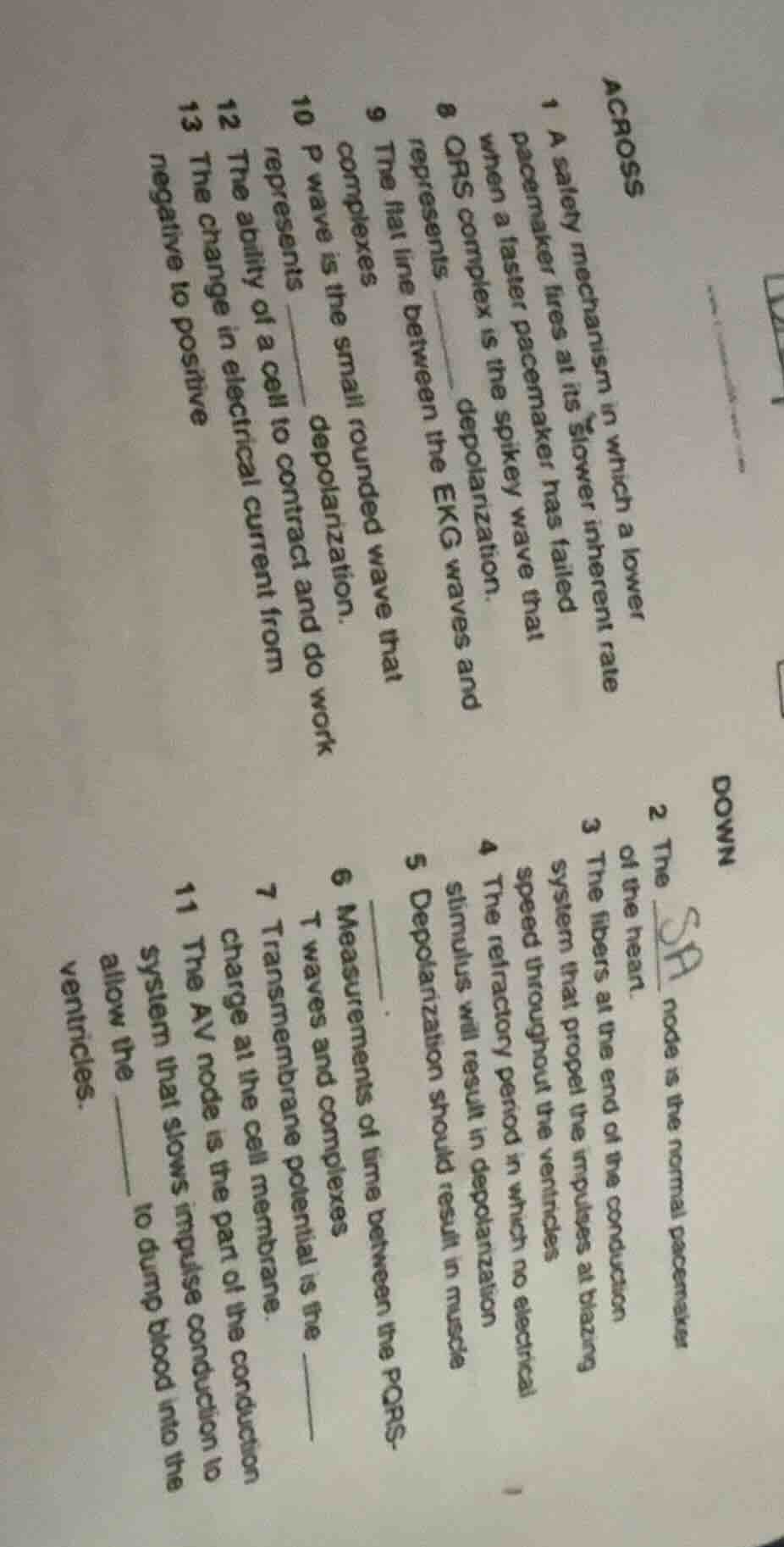
1 a safety mechanism in which a lower pacemaker fires at its slower inherent rate when a faster pacemaker has failed
8 qrs complex is the spikey wave that represents ____ depolarization.
9 the flat line between the ekg waves and complexes
10 p wave is the small rounded wave that represents ____ depolarization.
12 the ability of a cell to contract and do work
13 the change in electrical current from negative to positive
down
2 the ____ node is the normal pacemaker of the heart.
3 the fibers at the end of the conduction system that propel the impulses at blazing speed throughout the ventricles.
4 the refractory period in which no electrical stimulus will result in depolarization
5 depolarization should result in muscle ____.
6 measurements of time between the pqrst waves and complexes
7 transmembrane potential is the ____ charge at the cell membrane.
11 the av node is the part of the conduction system that slows impulse conduction to allow the ____ to dump blood into the ventricles.
These are fill-in-the-blank questions related to cardiac electrophysiology and EKG basics, key concepts in cardiac physiology.
- Across 1: This describes an escape rhythm, a backup pacemaker function.
- Across 8: The QRS complex corresponds to ventricular depolarization.
- Across 9: The flat line between EKG waves is the isoelectric line.
- Across 10: The P wave represents atrial depolarization.
- Across 12: Contractility is a cell's ability to contract and do work.
- Across 13: Depolarization is the shift from negative to positive electrical current.
- Down 2: The SA (Sinoatrial) node is the heart's normal pacemaker.
- Down 3: Purkinje fibers rapidly conduct impulses through the ventricles.
- Down 4: The absolute refractory period prevents any new depolarization.
- Down 5: Depolarization triggers muscle contraction.
- Down 6: Intervals are the time measurements between EKG waves/complexes.
- Down 7: Transmembrane potential is the electrical charge across the cell membrane.
- Down 11: The AV (Atrioventricular) node slows impulse conduction, and atrioventricular valves allow atrial blood to enter ventricles.

Snap & solve any problem in the app
Get step-by-step solutions on Sovi AI
Photo-based solutions with guided steps
Explore more problems and detailed explanations
Across:
- Escape Rhythm
- Ventricular
- Isoelectric Line
- Atrial
- Contractility
- Depolarization
Down:
- SA (Sinoatrial)
- Purkinje
- Absolute
- Contraction
- Intervals
- Electrical
- Atrioventricular; Atrioventricular Valves