QUESTION IMAGE
Question
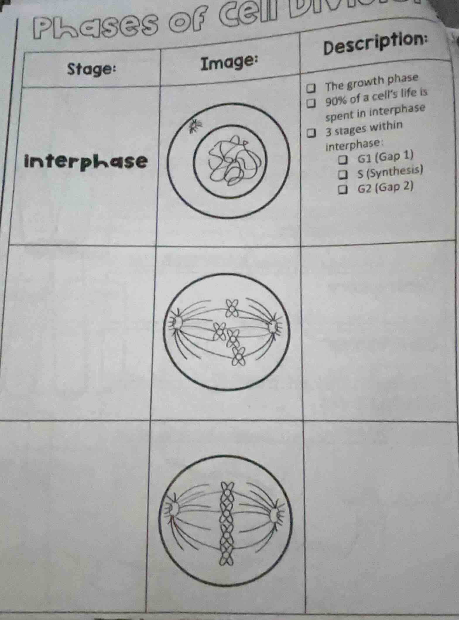
phases of cell division
stage: | image: | description:
interphase | image of interphase cell | the growth phase
90% of a cell’s life is spent in interphase
3 stages within interphase:
g1 (gap 1)
s (synthesis)
g2 (gap 2)
blank stage | image of cell with chromosomes aligned at equator |
blank stage | image of cell with chromosomes being pulled apart |
To determine the stages of cell division (mitosis) for the given images, we analyze each stage:
Second Row (Middle Image)
- Stage Identification: Metaphase (of mitosis).
- Reason: Chromosomes align at the cell’s equatorial plate (metaphase plate), and spindle fibers attach to centromeres. The image shows chromosomes lined up in the middle with spindle fibers from opposite poles.
Third Row (Bottom Image)
- Stage Identification: Anaphase (of mitosis).
- Reason: Sister chromatids separate and are pulled toward opposite poles by spindle fibers. The image shows chromatids moving apart, with spindle fibers shortening.
Filling the Table (assuming the task is to label the stages):
| Stage: | Image: | Description: |
|---|---|---|
| Metaphase | (Middle image) | Chromosomes align at metaphase plate; spindle fibers attach to centromeres. |
| Anaphase | (Bottom image) | Sister chromatids separate; pulled to opposite poles by spindle fibers. |
If the question was to identify the middle/bottom stages, the answers are:
- Middle stage: Metaphase
- Bottom stage: Anaphase

Snap & solve any problem in the app
Get step-by-step solutions on Sovi AI
Photo-based solutions with guided steps
Explore more problems and detailed explanations
To determine the stages of cell division (mitosis) for the given images, we analyze each stage:
Second Row (Middle Image)
- Stage Identification: Metaphase (of mitosis).
- Reason: Chromosomes align at the cell’s equatorial plate (metaphase plate), and spindle fibers attach to centromeres. The image shows chromosomes lined up in the middle with spindle fibers from opposite poles.
Third Row (Bottom Image)
- Stage Identification: Anaphase (of mitosis).
- Reason: Sister chromatids separate and are pulled toward opposite poles by spindle fibers. The image shows chromatids moving apart, with spindle fibers shortening.
Filling the Table (assuming the task is to label the stages):
| Stage: | Image: | Description: |
|---|---|---|
| Metaphase | (Middle image) | Chromosomes align at metaphase plate; spindle fibers attach to centromeres. |
| Anaphase | (Bottom image) | Sister chromatids separate; pulled to opposite poles by spindle fibers. |
If the question was to identify the middle/bottom stages, the answers are:
- Middle stage: Metaphase
- Bottom stage: Anaphase