QUESTION IMAGE
Question
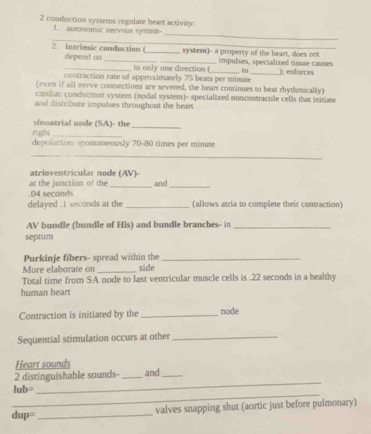
2 conduction systems regulate heart activity:
- autonomic nervous system-
- intrinsic conduction (____ system)- a property of the heart, does not depend on __ __ impulses, specialized tissue causes __ in only one direction (__ to ____); enforces contraction rate of approximately 75 beats per minute
(even if all nerve connections are severed, the heart continues to beat rhythmically)
cardiac conduction system (nodal system)- specialized noncontractile cells that initiate and distribute impulses throughout the heart
sinoatrial node (sa)- the ______
right ______
depolarizes spontaneously 70 - 80 times per minute
atrioventricular node (av)-
at the junction of the ____ and ____
.04 seconds
delayed.1 seconds at the ______ (allows atria to complete their contraction)
av bundle (bundle of his) and bundle branches- in ______
septum
purkinje fibers- spread within the ______
more elaborate on ______ side
total time from sa node to last ventricular muscle cells is.22 seconds in a healthy human heart
contraction is initiated by the ______ node
sequential stimulation occurs at other ______
heart sounds
2 distinguishable sounds- __ and __
lub=
dup= ______ valves snapping shut (aortic just before pulmonary)
Filling in the Blanks (Heart Conduction and Sounds)
1. Autonomic Nervous System
- The autonomic nervous system (sympathetic and parasympathetic divisions) regulates heart rate by sending neural impulses to the heart. It can speed up (sympathetic) or slow down (parasympathetic) the heart rate.
2. Intrinsic Conduction (Nodal) System
- Intrinsic conduction (nodal system) - a property of the heart, does not depend on external neural impulses. Specialized tissue causes impulse conduction in only one direction (atria to ventricles); enforces a contraction rate of approximately 75 beats per minute (even if all nerve connections are severed, the heart continues to beat rhythmically).
Sinoatrial Node (SA Node)
- Sinoatrial node (SA) - the pacemaker of the heart. Located in the right atrium. Depolarizes spontaneously 70 - 80 times per minute, initiating the heart’s electrical activity.
Atrioventricular Node (AV Node)
- Atrioventricular node (AV) - at the junction of the atria and ventricles. There is a 0.04 - second delay here, and an additional 0.1 - second delay at the AV node (allows atria to complete their contraction before ventricles contract).
AV Bundle (Bundle of His) and Bundle Branches
- AV bundle (bundle of His) and bundle branches - in the interventricular septum. They conduct impulses from the AV node to the ventricles.
Purkinje Fibers
- Purkinje fibers - spread within the ventricular myocardium (muscle of the ventricles). More elaborate on the left side (to ensure efficient contraction of the thicker left ventricle). Total time from SA node to last ventricular muscle cells is 0.22 seconds in a healthy human heart.
Contraction Initiation and Sequential Stimulation
- Contraction is initiated by the SA (sinoatrial) node. Sequential stimulation occurs at other nodes and conduction pathways (AV node, AV bundle, bundle branches, Purkinje fibers) to ensure coordinated contraction of the heart chambers.
Heart Sounds
- 2 distinguishable sounds - lub and dup.
- lub = closure of the atrioventricular (AV) valves (mitral and tricuspid valves) at the start of ventricular systole (contraction).
- dup = closure of the semilunar valves (aortic and pulmonary valves) at the start of ventricular diastole (relaxation). The aortic valve closes just before the pulmonary valve.
Final Answers (Filled Blanks)
- autonomic nervous system - sends neural impulses to regulate heart rate.
- intrinsic conduction (nodal system) - a property of the heart, does not depend on external neural impulses, specialized tissue causes impulse conduction in only one direction (atria to ventricles); enforces contraction rate of approximately 75 beats per minute
sinoatrial node (SA) - the pacemaker
right atrium
depolarizes spontaneously 70 - 80 times per minute
atrioventricular node (AV) -
at the junction of the atria and ventricles
.04 seconds
delayed.1 seconds at the AV node (allows atria to complete their contraction)
AV bundle (bundle of His) and bundle branches - in interventricular septum
Purkinje fibers - spread within the ventricular myocardium
More elaborate on left side
Contraction is initiated by the SA (sinoatrial) node
Sequential stimulation occurs at other nodes and conduction pathways
Heart sounds
2 distinguishable sounds - lub and dup
lub = closure of the atrioventricular (AV) valves
dup = closure of the semilunar valves snapp…

Snap & solve any problem in the app
Get step-by-step solutions on Sovi AI
Photo-based solutions with guided steps
Explore more problems and detailed explanations
Filling in the Blanks (Heart Conduction and Sounds)
1. Autonomic Nervous System
- The autonomic nervous system (sympathetic and parasympathetic divisions) regulates heart rate by sending neural impulses to the heart. It can speed up (sympathetic) or slow down (parasympathetic) the heart rate.
2. Intrinsic Conduction (Nodal) System
- Intrinsic conduction (nodal system) - a property of the heart, does not depend on external neural impulses. Specialized tissue causes impulse conduction in only one direction (atria to ventricles); enforces a contraction rate of approximately 75 beats per minute (even if all nerve connections are severed, the heart continues to beat rhythmically).
Sinoatrial Node (SA Node)
- Sinoatrial node (SA) - the pacemaker of the heart. Located in the right atrium. Depolarizes spontaneously 70 - 80 times per minute, initiating the heart’s electrical activity.
Atrioventricular Node (AV Node)
- Atrioventricular node (AV) - at the junction of the atria and ventricles. There is a 0.04 - second delay here, and an additional 0.1 - second delay at the AV node (allows atria to complete their contraction before ventricles contract).
AV Bundle (Bundle of His) and Bundle Branches
- AV bundle (bundle of His) and bundle branches - in the interventricular septum. They conduct impulses from the AV node to the ventricles.
Purkinje Fibers
- Purkinje fibers - spread within the ventricular myocardium (muscle of the ventricles). More elaborate on the left side (to ensure efficient contraction of the thicker left ventricle). Total time from SA node to last ventricular muscle cells is 0.22 seconds in a healthy human heart.
Contraction Initiation and Sequential Stimulation
- Contraction is initiated by the SA (sinoatrial) node. Sequential stimulation occurs at other nodes and conduction pathways (AV node, AV bundle, bundle branches, Purkinje fibers) to ensure coordinated contraction of the heart chambers.
Heart Sounds
- 2 distinguishable sounds - lub and dup.
- lub = closure of the atrioventricular (AV) valves (mitral and tricuspid valves) at the start of ventricular systole (contraction).
- dup = closure of the semilunar valves (aortic and pulmonary valves) at the start of ventricular diastole (relaxation). The aortic valve closes just before the pulmonary valve.
Final Answers (Filled Blanks)
- autonomic nervous system - sends neural impulses to regulate heart rate.
- intrinsic conduction (nodal system) - a property of the heart, does not depend on external neural impulses, specialized tissue causes impulse conduction in only one direction (atria to ventricles); enforces contraction rate of approximately 75 beats per minute
sinoatrial node (SA) - the pacemaker
right atrium
depolarizes spontaneously 70 - 80 times per minute
atrioventricular node (AV) -
at the junction of the atria and ventricles
.04 seconds
delayed.1 seconds at the AV node (allows atria to complete their contraction)
AV bundle (bundle of His) and bundle branches - in interventricular septum
Purkinje fibers - spread within the ventricular myocardium
More elaborate on left side
Contraction is initiated by the SA (sinoatrial) node
Sequential stimulation occurs at other nodes and conduction pathways
Heart sounds
2 distinguishable sounds - lub and dup
lub = closure of the atrioventricular (AV) valves
dup = closure of the semilunar valves snapping shut (aortic just before pulmonary)