QUESTION IMAGE
Question
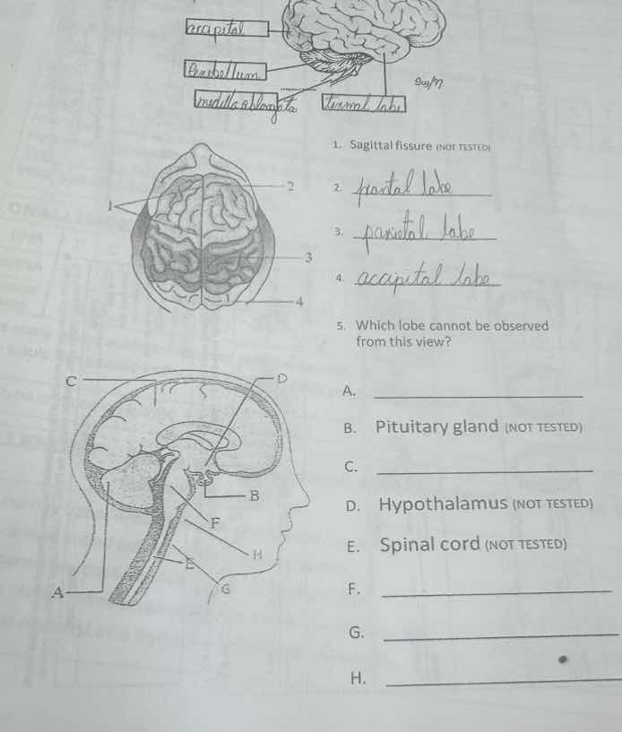
- sagittal fissure (not tested)
2.
3.
4.
- which lobe cannot be observed from this view?
a.
b. pituitary gland (not tested)
c.
d. hypothalamus (not tested)
e. spinal cord (not tested)
f.
g.
h.
To solve these questions, we analyze the brain structures and lobes based on the given views (dorsal/top view and sagittal view):
Question 2, 3, 4 (Labeling Lobes in Dorsal View):
In a dorsal (top) view of the brain, the visible lobes are:
- 2. Frontal Lobe: Anterior (front) part of the cerebrum.
- 3. Parietal Lobe: Middle (between frontal and occipital) part.
- 4. Occipital Lobe: Posterior (back) part.
Question 5 (Lobe Not Visible in Dorsal View):
The dorsal view shows the top of the brain. The Temporal Lobe is located on the side (lateral) of the brain, so it cannot be seen from a top-down (dorsal) view.
Labeling Structures in Sagittal View:
In a sagittal (side) view of the brain:
- A. Cerebellum: Posterior, below the cerebrum (looks like a “bulb” at the back/bottom of the brain).
- C. Cerebrum (Cerebral Cortex): Large, outer part of the brain (front/top in sagittal view).
- F. Pons: Structure between the midbrain and medulla (part of the brainstem).
- G. Medulla Oblongata: Inferior part of the brainstem (connects to spinal cord).
- H. Spinal Cord (wait, no—H is likely the brainstem? Wait, correction: In sagittal view, the brainstem includes midbrain, pons, medulla. Let’s recheck:
- A: Cerebellum (correct, posterior, rounded).
- C: Cerebrum (cerebral cortex, correct).
- F: Pons (correct, between midbrain and medulla).
- G: Medulla Oblongata (correct, inferior brainstem).
- H: Maybe Midbrain? Wait, the labels: H is likely the Midbrain or Brainstem? Alternatively, H could be the spinal cord (but E is labeled spinal cord). Let’s align with standard sagittal brain anatomy:
- Cerebellum (A), Cerebrum (C), Pons (F), Medulla Oblongata (G), Spinal Cord (E), Hypothalamus (D), Pituitary (B), etc.
Final Answers:
- $\boldsymbol{\text{Frontal Lobe}}$
- $\boldsymbol{\text{Parietal Lobe}}$
- $\boldsymbol{\text{Occipital Lobe}}$
- A. $\boldsymbol{\text{Temporal Lobe}}$
A (structure): $\boldsymbol{\text{Cerebellum}}$
C (structure): $\boldsymbol{\text{Cerebrum (Cerebral Cortex)}}$
F (structure): $\boldsymbol{\text{Pons}}$
G (structure): $\boldsymbol{\text{Medulla Oblongata}}$
H (structure): $\boldsymbol{\text{Midbrain}}$ (or $\boldsymbol{\text{Brainstem}}$; adjust based on diagram)
(Note: Some labels may vary slightly based on the diagram’s specific markings, but the key is identifying lobes and brain structures by their location.)

Snap & solve any problem in the app
Get step-by-step solutions on Sovi AI
Photo-based solutions with guided steps
Explore more problems and detailed explanations
To solve these questions, we analyze the brain structures and lobes based on the given views (dorsal/top view and sagittal view):
Question 2, 3, 4 (Labeling Lobes in Dorsal View):
In a dorsal (top) view of the brain, the visible lobes are:
- 2. Frontal Lobe: Anterior (front) part of the cerebrum.
- 3. Parietal Lobe: Middle (between frontal and occipital) part.
- 4. Occipital Lobe: Posterior (back) part.
Question 5 (Lobe Not Visible in Dorsal View):
The dorsal view shows the top of the brain. The Temporal Lobe is located on the side (lateral) of the brain, so it cannot be seen from a top-down (dorsal) view.
Labeling Structures in Sagittal View:
In a sagittal (side) view of the brain:
- A. Cerebellum: Posterior, below the cerebrum (looks like a “bulb” at the back/bottom of the brain).
- C. Cerebrum (Cerebral Cortex): Large, outer part of the brain (front/top in sagittal view).
- F. Pons: Structure between the midbrain and medulla (part of the brainstem).
- G. Medulla Oblongata: Inferior part of the brainstem (connects to spinal cord).
- H. Spinal Cord (wait, no—H is likely the brainstem? Wait, correction: In sagittal view, the brainstem includes midbrain, pons, medulla. Let’s recheck:
- A: Cerebellum (correct, posterior, rounded).
- C: Cerebrum (cerebral cortex, correct).
- F: Pons (correct, between midbrain and medulla).
- G: Medulla Oblongata (correct, inferior brainstem).
- H: Maybe Midbrain? Wait, the labels: H is likely the Midbrain or Brainstem? Alternatively, H could be the spinal cord (but E is labeled spinal cord). Let’s align with standard sagittal brain anatomy:
- Cerebellum (A), Cerebrum (C), Pons (F), Medulla Oblongata (G), Spinal Cord (E), Hypothalamus (D), Pituitary (B), etc.
Final Answers:
- $\boldsymbol{\text{Frontal Lobe}}$
- $\boldsymbol{\text{Parietal Lobe}}$
- $\boldsymbol{\text{Occipital Lobe}}$
- A. $\boldsymbol{\text{Temporal Lobe}}$
A (structure): $\boldsymbol{\text{Cerebellum}}$
C (structure): $\boldsymbol{\text{Cerebrum (Cerebral Cortex)}}$
F (structure): $\boldsymbol{\text{Pons}}$
G (structure): $\boldsymbol{\text{Medulla Oblongata}}$
H (structure): $\boldsymbol{\text{Midbrain}}$ (or $\boldsymbol{\text{Brainstem}}$; adjust based on diagram)
(Note: Some labels may vary slightly based on the diagram’s specific markings, but the key is identifying lobes and brain structures by their location.)