QUESTION IMAGE
Question
sports medicine
worksheet 2
date:
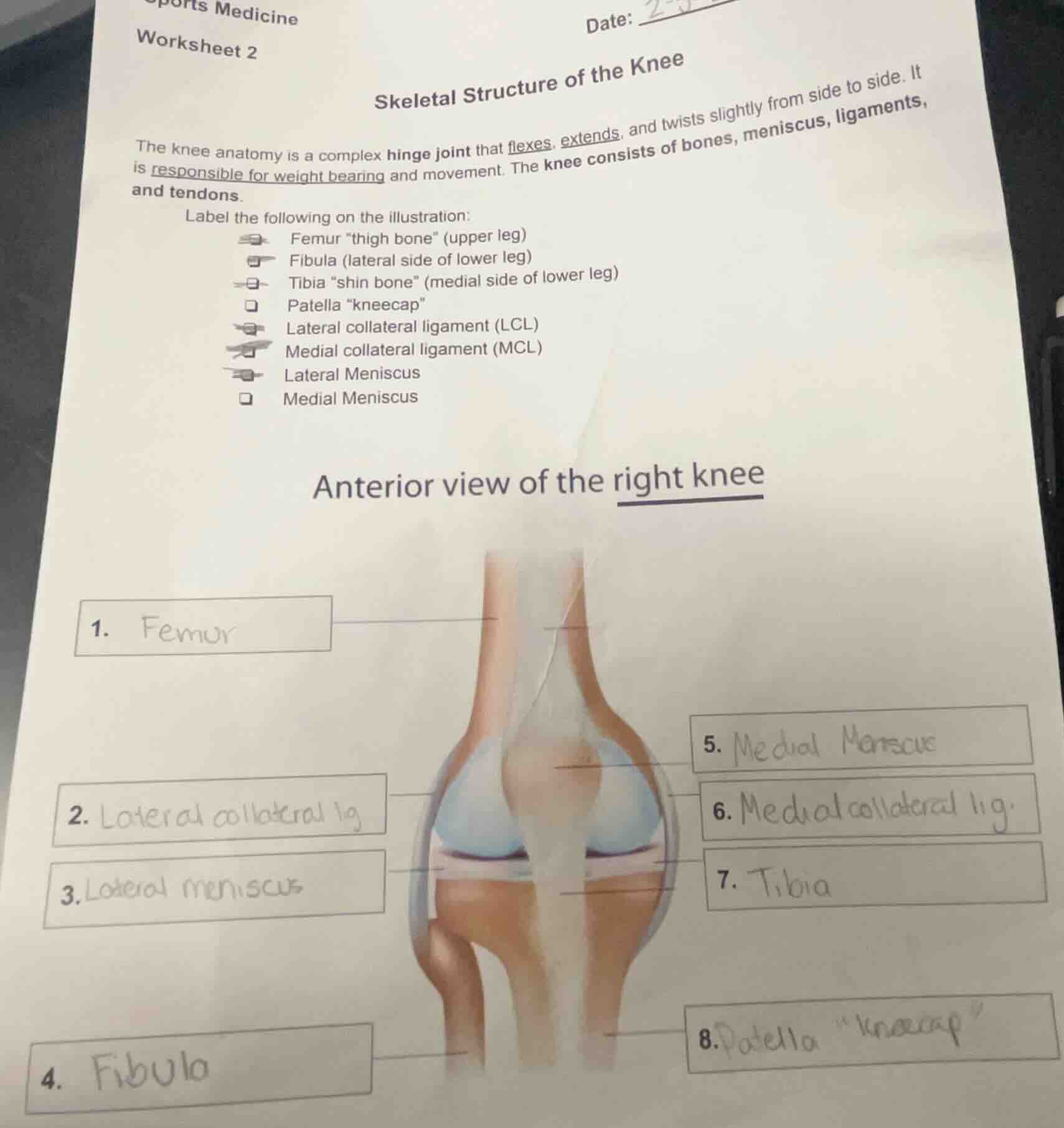
skeletal structure of the knee
the knee anatomy is a complex hinge joint that flexes, extends, and twists slightly from side to side. it is responsible for weight bearing and movement. the knee consists of bones, meniscus, ligaments, and tendons.
label the following on the illustration:
femur “thigh bone” (upper leg)
fibula (lateral side of lower leg)
tibia “shin bone” (medial side of lower leg)
patella “kneecap”
lateral collateral ligament (lcl)
medial collateral ligament (mcl)
lateral meniscus
medial meniscus
anterior view of the right knee
- femur
- lateral collateral lig
- lateral meniscus
- fibula
- medial meniscus
- medial collateral lig.
- tibia
- patella “kneecap”
The worksheet is about the skeletal structure of the knee, which falls under the subfield of Biology (specifically Anatomy, a part of Biology) as it deals with the anatomical structure of the knee joint, including bones, ligaments, meniscus, etc. The task is to label the parts, and the provided labels match the anatomical terms: 1 is Femur (thigh bone), 2 is Lateral collateral ligament (LCL), 3 is Lateral Meniscus, 4 is Fibula, 5 is Medial Meniscus, 6 is Medial collateral ligament (MCL), 7 is Tibia (shin bone), 8 is Patella (kneecap). These labels correctly identify the respective structures in the anterior view of the right knee.

Snap & solve any problem in the app
Get step-by-step solutions on Sovi AI
Photo-based solutions with guided steps
Explore more problems and detailed explanations
- Femur (thigh bone)
- Lateral collateral ligament (LCL)
- Lateral Meniscus
- Fibula
- Medial Meniscus
- Medial collateral ligament (MCL)
- Tibia (shin bone)
- Patella (kneecap)