QUESTION IMAGE
Question
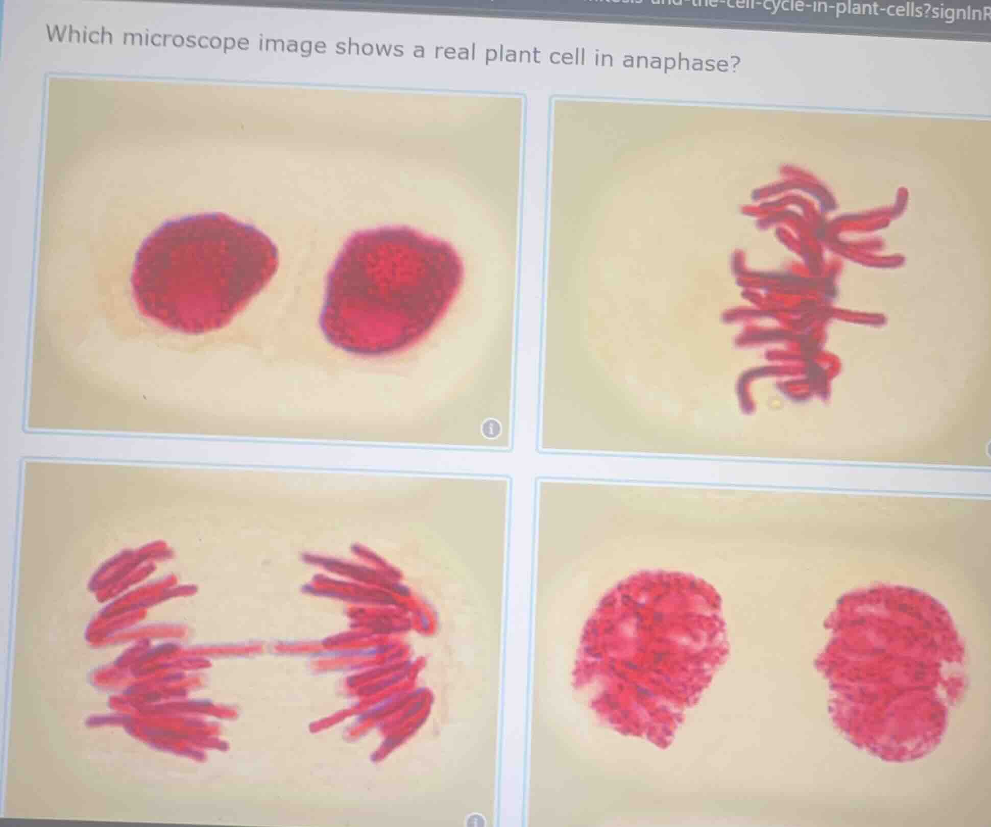
which microscope image shows a real plant cell in anaphase?
Brief Explanations
Anaphase of mitosis in plant cells is defined by sister chromatids separating and being pulled to opposite poles of the cell, creating two distinct groups of chromosomes moving apart.
- The top-left image shows two fully formed nuclei, which is telophase/cytokinesis.
- The top-right image shows chromosomes aligned at the cell equator, which is metaphase.
- The bottom-left image shows chromosomes pulled into two separate groups moving toward opposite poles, matching anaphase.
- The bottom-right image shows cells with condensed chromosomes that have not begun separating, which is prophase or early metaphase.

Snap & solve any problem in the app
Get step-by-step solutions on Sovi AI
Photo-based solutions with guided steps
Explore more problems and detailed explanations
Bottom-left image (the one with two distinct groups of chromosomes being pulled to opposite ends of the cell)