QUESTION IMAGE
Question
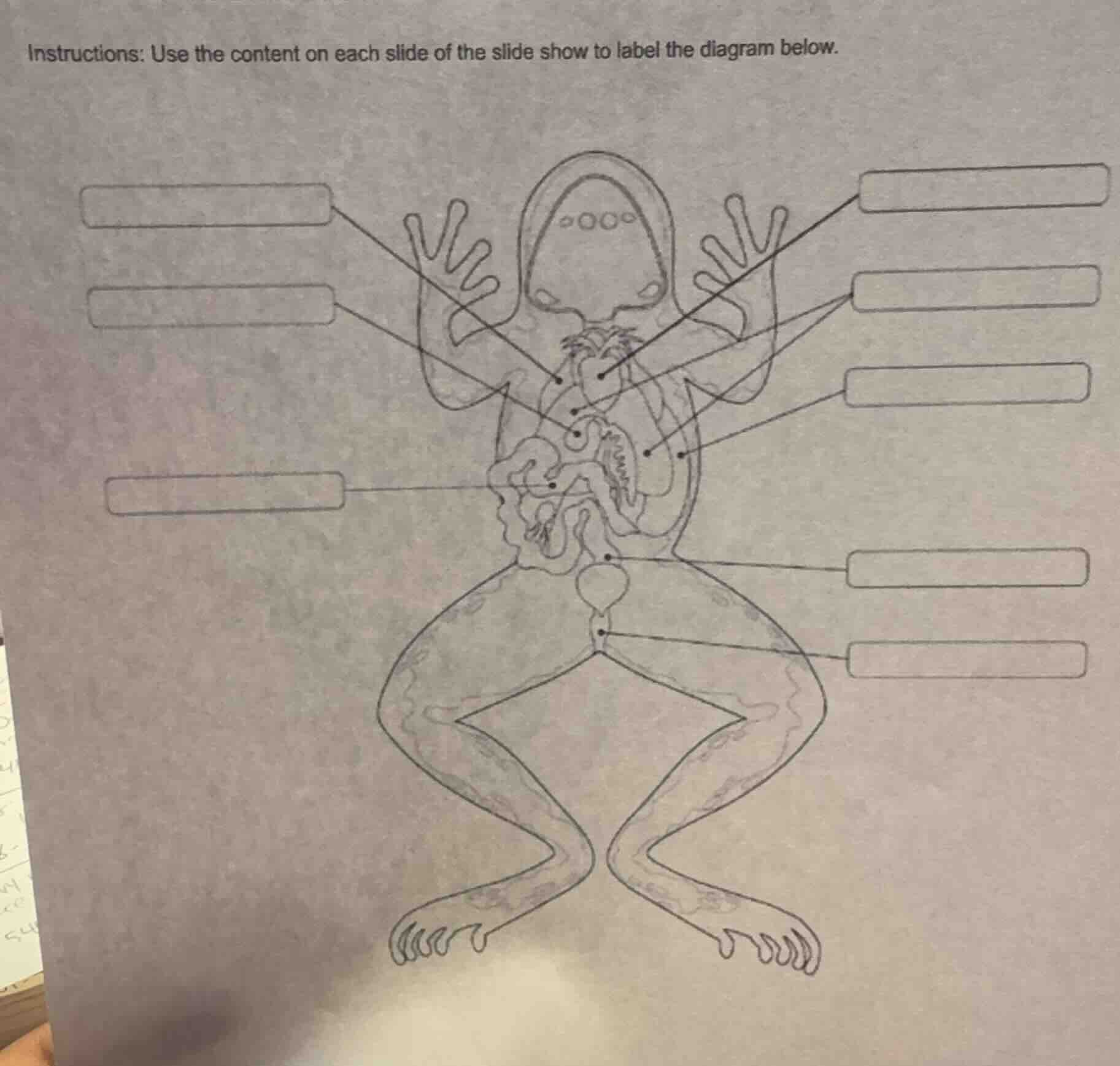
instructions: use the content on each slide of the slide show to label the diagram below.
To label the frog's anatomy diagram, we typically identify structures like the heart, lungs, liver, stomach, intestines, bladder, cloaca, and other organs. However, since the slide show content (which has the specific labels) isn't provided here, we can't fill in the exact labels. But generally, for a frog's internal anatomy:
- Heart: Central, muscular organ for circulation.
- Lungs: Near the heart, for respiration.
- Liver: Large, lobed organ (often multiple lobes) near the stomach.
- Stomach: Sac-like organ for digestion.
- Small Intestine: Coiled tube for nutrient absorption.
- Large Intestine/Rectum: Shorter, for waste processing.
- Bladder: Stores urine, near the cloaca.
- Cloaca: Common opening for waste and reproduction.
If the slide show includes terms like heart, lung, liver, stomach, small intestine, large intestine, urinary bladder, cloaca, you would match each line to the corresponding structure based on their typical positions (e.g., the heart is central, lungs are above/around it, liver is below the heart, stomach is below the liver, intestines are coiled, bladder is below the intestines, cloaca is at the bottom).
Since the slide content is missing, you’ll need to refer to the slide show to get the exact labels and place them in the correct boxes based on the diagram’s structure (e.g., top-left lines might point to heart/lung, middle lines to digestive organs, bottom lines to bladder/cloaca).

Snap & solve any problem in the app
Get step-by-step solutions on Sovi AI
Photo-based solutions with guided steps
Explore more problems and detailed explanations
To label the frog's anatomy diagram, we typically identify structures like the heart, lungs, liver, stomach, intestines, bladder, cloaca, and other organs. However, since the slide show content (which has the specific labels) isn't provided here, we can't fill in the exact labels. But generally, for a frog's internal anatomy:
- Heart: Central, muscular organ for circulation.
- Lungs: Near the heart, for respiration.
- Liver: Large, lobed organ (often multiple lobes) near the stomach.
- Stomach: Sac-like organ for digestion.
- Small Intestine: Coiled tube for nutrient absorption.
- Large Intestine/Rectum: Shorter, for waste processing.
- Bladder: Stores urine, near the cloaca.
- Cloaca: Common opening for waste and reproduction.
If the slide show includes terms like heart, lung, liver, stomach, small intestine, large intestine, urinary bladder, cloaca, you would match each line to the corresponding structure based on their typical positions (e.g., the heart is central, lungs are above/around it, liver is below the heart, stomach is below the liver, intestines are coiled, bladder is below the intestines, cloaca is at the bottom).
Since the slide content is missing, you’ll need to refer to the slide show to get the exact labels and place them in the correct boxes based on the diagram’s structure (e.g., top-left lines might point to heart/lung, middle lines to digestive organs, bottom lines to bladder/cloaca).