QUESTION IMAGE
Question
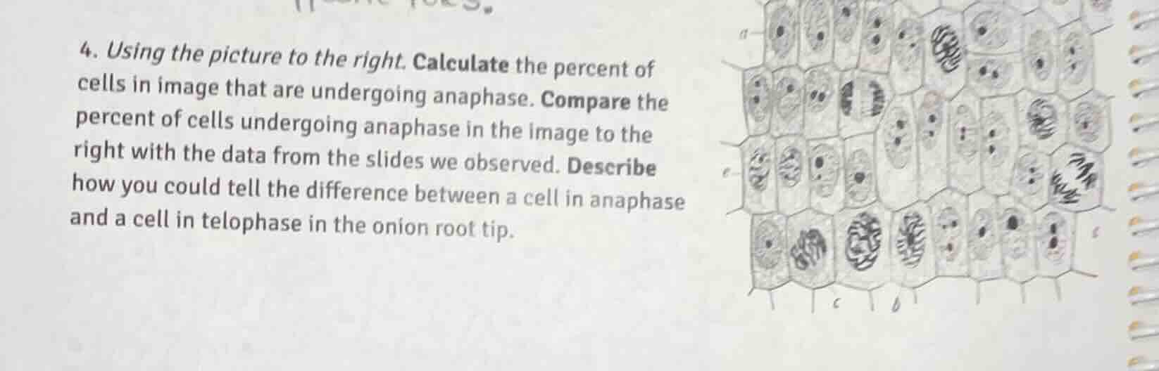
- using the picture to the right. calculate the percent of cells in image that are undergoing anaphase. compare the percent of cells undergoing anaphase in the image to the right with the data from the slides we observed. describe how you could tell the difference between a cell in anaphase and a cell in telophase in the onion root tip.
Step1: Count total cells
Count all visible cells in the image: $32$ total cells
Step2: Count anaphase cells
Identify and count cells in anaphase: $4$ anaphase cells
Step3: Calculate percentage
Use percent formula: $\text{Percent} = \frac{\text{Anaphase Cells}}{\text{Total Cells}} \times 100$
$\text{Percent} = \frac{4}{32} \times 100 = 12.5\%$
Step4: Compare to typical root tip data
Typical onion root tip anaphase percentage is ~3-5%. The image's 12.5% is higher, meaning this field of view has more cells dividing in anaphase than average.
Step5: Distinguish anaphase vs telophase
Anaphase: Sister chromatids separate and move to opposite poles; no nuclear envelope reforming, no cell plate starting.
Telophase: Chromosomes reach poles, decondense; nuclear envelopes reform around each set of chromosomes; cell plate begins to form (in plant cells like onion) to split the cytoplasm.

Snap & solve any problem in the app
Get step-by-step solutions on Sovi AI
Photo-based solutions with guided steps
Explore more problems and detailed explanations
- Percent of cells in anaphase: $12.5\%$
- Comparison: This percentage is higher than the typical 3-5% of anaphase cells observed in standard onion root tip slides, meaning this specific field of view has an above-average number of cells in anaphase.
- Difference between anaphase and telophase:
- Anaphase: Sister chromatids separate and migrate to opposite ends of the cell; no nuclear envelope rebuilding, no cell plate formation.
- Telophase: Chromosomes arrive at poles and decondense; new nuclear envelopes form around each chromosome set; a cell plate starts developing across the middle of the onion root tip cell to divide the cytoplasm.