QUESTION IMAGE
Question
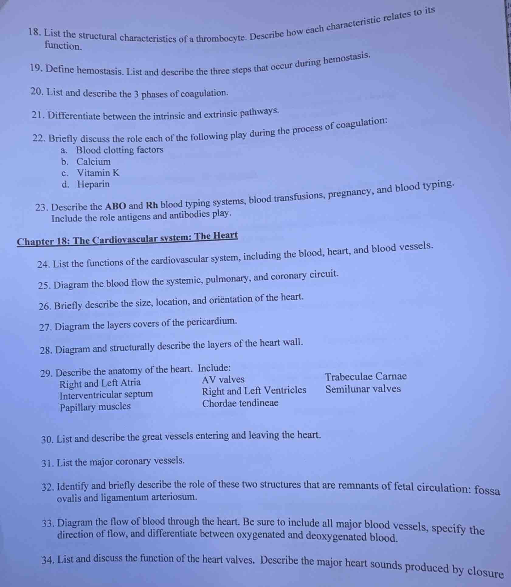
- list the structural characteristics of a thrombocyte. describe how each characteristic relates to its function.
- define hemostasis. list and describe the three steps that occur during hemostasis.
- list and describe the 3 phases of coagulation.
- differentiate between the intrinsic and extrinsic pathways.
- briefly discuss the role each of the following play during the process of coagulation:
a. blood clotting factors
b. calcium
c. vitamin k
d. heparin
- describe the abo and rh blood typing systems, blood transfusions, pregnancy, and blood typing. include the role antigens and antibodies play.
chapter 18: the cardiovascular system: the heart
- list the functions of the cardiovascular system, including the blood, heart, and blood vessels.
- diagram the blood flow the systemic, pulmonary, and coronary circuit.
- briefly describe the size, location, and orientation of the heart.
- diagram the layers covers of the pericardium.
- diagram and structurally describe the layers of the heart wall.
- describe the anatomy of the heart. include:
right and left atria
interventricular septum
papillary muscles
av valves
right and left ventricles
chordae tendineae
trabeculae carnae
semilunar valves
- list and describe the great vessels entering and leaving the heart.
- list the major coronary vessels.
- identify and briefly describe the role of these two structures that are remnants of fetal circulation: fossa ovalis and ligamentum arteriosum.
- diagram the flow of blood through the heart. be sure to include all major blood vessels, specify the direction of flow, and differentiate between oxygenated and deoxygenated blood.
- list and discuss the function of the heart valves. describe the major heart sounds produced by closure
Question 18
Thrombocytes (platelets) are small, anucleate cell fragments. Their structural features support blood clotting:
- Small, disc shape: Lets them easily move through narrow blood vessels and accumulate at injury sites.
- Glycoprotein surface receptors: Bind to damaged vessel walls and other platelets to form clots.
- Cytoplasmic granules (alpha, dense): Store clotting factors, calcium, and signaling molecules (like ADP) that trigger platelet activation and aggregation.
- Contractile proteins (actin, myosin): Allow platelets to change shape (spreading) when activated, and shrink clots to stop bleeding.
Question 19
Hemostasis: The body's process to stop bleeding from damaged blood vessels.
- Vascular spasm: Damaged vessels constrict, reducing blood flow to the injury. Triggered by vessel injury and platelet signaling.
- Platelet plug formation: Platelets stick to damaged vessel collagen, activate (change shape, release granules), and clump together to form a temporary plug.
- Coagulation (blood clotting): Fibrin proteins form a mesh that traps blood cells and platelets, creating a stable clot to seal the injury.
Question 20
The three phases of coagulation:
- Activation of prothrombinase: A cascade of reactions (intrinsic or extrinsic pathway) activates the enzyme prothrombinase.
- Prothrombin conversion: Prothrombinase converts prothrombin (a plasma protein) into thrombin (an active enzyme).
- Fibrin formation: Thrombin converts soluble fibrinogen into insoluble fibrin threads, which cross-link to form a clot mesh.
Question 21
| Feature | Intrinsic Pathway | Extrinsic Pathway |
|---|---|---|
| Reaction Speed | Slow (several minutes) | Fast (seconds) |
| Factors Involved | Factors XII, XI, IX, VIII | Tissue factor (Factor III), Factor VII |
| Location | Starts inside the bloodstream | Starts outside the bloodstream (tissue) |
Question 22
a. Blood clotting factors: Plasma proteins (numbered I-XIII) that act in a cascade; each activated factor triggers the next, leading to fibrin clot formation.
b. Calcium: Required as a cofactor for multiple steps in both intrinsic and extrinsic coagulation pathways to activate clotting factors.
c. Vitamin K: Necessary for the liver to produce functional clotting factors II, VII, IX, and X (deficiency leads to bleeding disorders).
d. Heparin: An anticoagulant that inhibits thrombin activity, preventing fibrin formation and stopping unwanted clotting.
Question 23
- ABO System: Based on antigens (A, B) on red blood cells (RBCs):
- Type A: A antigens on RBCs, anti-B antibodies in plasma
- Type B: B antigens on RBCs, anti-A antibodies in plasma
- Type AB: A + B antigens, no anti-A/B antibodies (universal recipient)
- Type O: No A/B antigens, anti-A + anti-B antibodies (universal donor)
- Transfusion risk: Antibodies in recipient plasma attack mismatched antigens on donor RBCs, causing RBC rupture.
- Rh System: Based on the Rh(D) antigen on RBCs:
- Rh-positive: Rh antigen present; Rh-negative: no Rh antigen, no natural anti-Rh antibodies.
- Pregnancy risk: If Rh-negative mother carries Rh-positive fetus, mother may develop anti-Rh antibodies. In subsequent Rh-positive pregnancies, these antibodies can attack fetal RBCs (hemolytic disease of the newborn).
- Blood typing: Tests for presence of A/B/Rh antige…

Snap & solve any problem in the app
Get step-by-step solutions on Sovi AI
Photo-based solutions with guided steps
Explore more problems and detailed explanations
All questions 18-34 are answered above in the corresponding sections.