QUESTION IMAGE
Question
4.
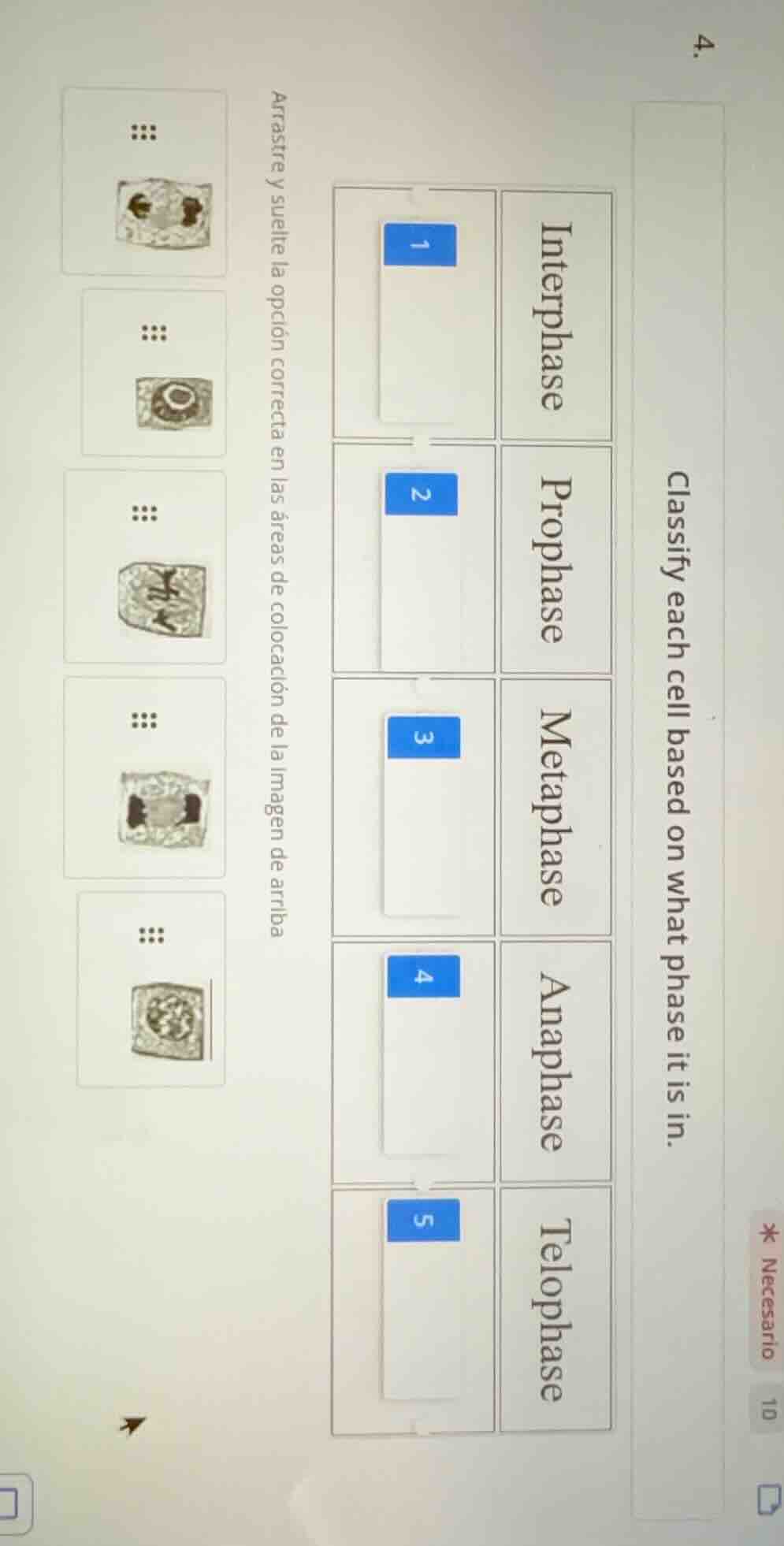
classify each cell based on what phase it is in.
interphase
1
prophase
2
metaphase
3
anaphase
4
telophase
5
arrastre y suelte la opción correcta en las áreas de colocación de la imagen de arriba
To solve this, we analyze each cell image and match it to the correct mitotic phase:
Step 1: Analyze Cell 1 (Top - Interphase)
Interphase cells have a nucleus with visible chromatin (not condensed chromosomes). The 5th cell image (bottom - nucleus with dispersed chromatin) matches Interphase. So, drag cell 5 to Interphase (1).
Step 2: Analyze Cell 2 (Prophase)
Prophase: Chromosomes condense, nucleus/nucleolus start to break down. The 2nd cell image (nucleus with condensed chromosomes, nucleolus visible) matches Prophase. Drag cell 2 to Prophase (2).
Step 3: Analyze Cell 3 (Metaphase)
Metaphase: Chromosomes align at the cell equator. The 3rd cell image (chromosomes lined up in the middle) matches Metaphase. Drag cell 3 to Metaphase (3).
Step 4: Analyze Cell 4 (Anaphase)
Anaphase: Sister chromatids separate and move to opposite poles. The 4th cell image (chromatids pulled apart) matches Anaphase. Drag cell 4 to Anaphase (4).
Step 5: Analyze Cell 5 (Telophase)
Telophase: Nuclear envelopes reform, chromosomes decondense. The 1st cell image (two new nuclei forming) matches Telophase. Drag cell 1 to Telophase (5).
Final Matching:
- Interphase → Cell 5
- Prophase → Cell 2
- Metaphase → Cell 3
- Anaphase → Cell 4
- Telophase → Cell 1
(Note: The exact image details confirm these matches based on mitotic phase characteristics.)

Snap & solve any problem in the app
Get step-by-step solutions on Sovi AI
Photo-based solutions with guided steps
Explore more problems and detailed explanations
To solve this, we analyze each cell image and match it to the correct mitotic phase:
Step 1: Analyze Cell 1 (Top - Interphase)
Interphase cells have a nucleus with visible chromatin (not condensed chromosomes). The 5th cell image (bottom - nucleus with dispersed chromatin) matches Interphase. So, drag cell 5 to Interphase (1).
Step 2: Analyze Cell 2 (Prophase)
Prophase: Chromosomes condense, nucleus/nucleolus start to break down. The 2nd cell image (nucleus with condensed chromosomes, nucleolus visible) matches Prophase. Drag cell 2 to Prophase (2).
Step 3: Analyze Cell 3 (Metaphase)
Metaphase: Chromosomes align at the cell equator. The 3rd cell image (chromosomes lined up in the middle) matches Metaphase. Drag cell 3 to Metaphase (3).
Step 4: Analyze Cell 4 (Anaphase)
Anaphase: Sister chromatids separate and move to opposite poles. The 4th cell image (chromatids pulled apart) matches Anaphase. Drag cell 4 to Anaphase (4).
Step 5: Analyze Cell 5 (Telophase)
Telophase: Nuclear envelopes reform, chromosomes decondense. The 1st cell image (two new nuclei forming) matches Telophase. Drag cell 1 to Telophase (5).
Final Matching:
- Interphase → Cell 5
- Prophase → Cell 2
- Metaphase → Cell 3
- Anaphase → Cell 4
- Telophase → Cell 1
(Note: The exact image details confirm these matches based on mitotic phase characteristics.)