QUESTION IMAGE
Question
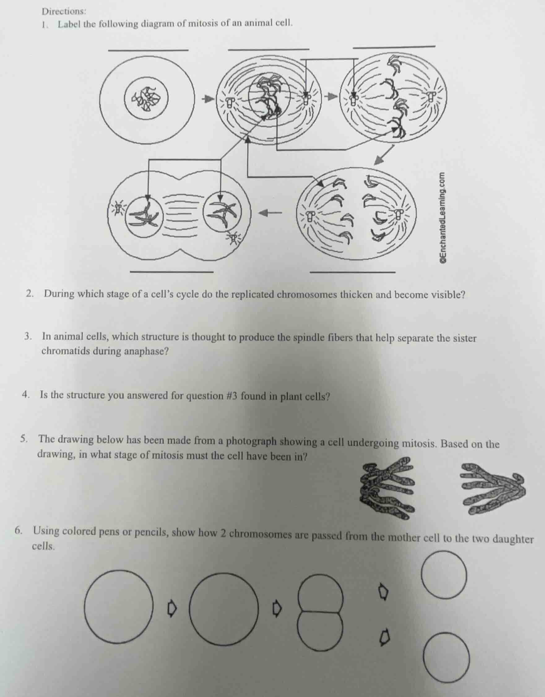
directions:
- label the following diagram of mitosis of an animal cell.
- during which stage of a cell’s cycle do the replicated chromosomes thicken and become visible?
- in animal cells, which structure is thought to produce the spindle fibers that help separate the sister chromatids during anaphase?
- is the structure you answered for question #3 found in plant cells?
- the drawing below has been made from a photograph showing a cell undergoing mitosis. based on the drawing, in what stage of mitosis must the cell have been in?
- using colored pens or pencils, show how 2 chromosomes are passed from the mother cell to the two daughter cells.
Brief Explanations
- Labels the mitosis stages and key structures based on animal cell mitosis characteristics.
- Identifies the stage where chromosomes condense into visible structures.
- Names the animal cell structure that generates spindle fibers.
- Compares presence of the structure between animal and plant cells.
- Identifies mitosis stage by aligned, paired chromosomes at the cell equator.
- Outlines the path of 2 chromosomes from parent to daughter cells during mitosis.

Snap & solve any problem in the app
Get step-by-step solutions on Sovi AI
Photo-based solutions with guided steps
Explore more problems and detailed explanations
- Top left (stage): Prophase
Middle top (stage): Metaphase; (structure, center): Chromosome (with sister chromatids); (structure, poles): Centriole
Top right (stage): Anaphase
Bottom right (stage): Telophase
Bottom left (stage): Cytokinesis
- Prophase
- Centriole (centrosome)
- No, plant cells do not have centrioles.
- Metaphase
- - Color the two diamond-shaped chromosomes (representing the mother cell's chromosomes) with two distinct colors.
- Copy both colored chromosomes into the second circle (cell in prophase/metaphase, with chromosomes replicated, so each diamond becomes a pair of identical colored diamonds attached at a center point).
- Draw one of each colored single diamond chromosome into each half of the dividing cell (anaphase/telophase).
- Draw one of each colored single diamond chromosome into each of the two final daughter cells.