QUESTION IMAGE
Question
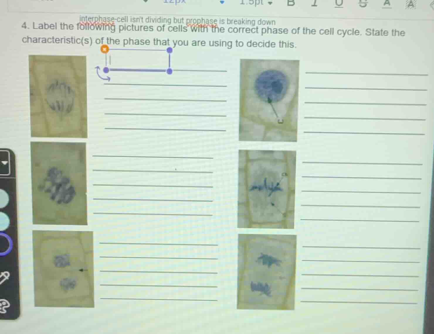
- label the following pictures of cells with the correct phase of the cell cycle. state the characteristic(s) of the phase that you are using to decide this.
To solve this, we analyze each cell image based on mitotic phase characteristics:
Top - Left Cell (First Row, First Column)
- Phase: Anaphase
- Characteristic: Chromosomes are pulled apart to opposite poles of the cell (sister chromatids separate and move towards spindle poles).
Top - Right Cell (First Row, Second Column)
- Phase: Interphase
- Characteristic: The nucleus is intact, chromatin is diffuse (no visible chromosomes), and the cell is in a non - dividing state (preparing for division or in a resting state).
Middle - Left Cell (Second Row, First Column)
- Phase: Prophase
- Characteristic: Chromosomes are condensing (becoming visible and coiled), and the nuclear envelope may be starting to break down.
Middle - Right Cell (Second Row, Second Column)
- Phase: Metaphase
- Characteristic: Chromosomes are aligned along the equatorial plate (middle) of the cell, attached to spindle fibers.
Bottom - Left Cell (Third Row, First Column)
- Phase: Telophase
- Characteristic: Two distinct nuclei are forming (cell is beginning to divide into two daughter cells), and a cell plate or cleavage furrow may be starting to appear (in plant cells, a cell plate forms).
Bottom - Right Cell (Third Row, Second Column)
- Phase: Anaphase (or Late Anaphase)
- Characteristic: Chromosomes are separated and moving towards opposite ends of the cell (similar to the top - left cell, but may be at a slightly later stage of anaphase).
(Note: The exact phase identification depends on the precise visual details of chromosome arrangement, nuclear envelope status, and cell structure in each image. The above is a general guide based on typical mitotic phase features.)

Snap & solve any problem in the app
Get step-by-step solutions on Sovi AI
Photo-based solutions with guided steps
Explore more problems and detailed explanations
To solve this, we analyze each cell image based on mitotic phase characteristics:
Top - Left Cell (First Row, First Column)
- Phase: Anaphase
- Characteristic: Chromosomes are pulled apart to opposite poles of the cell (sister chromatids separate and move towards spindle poles).
Top - Right Cell (First Row, Second Column)
- Phase: Interphase
- Characteristic: The nucleus is intact, chromatin is diffuse (no visible chromosomes), and the cell is in a non - dividing state (preparing for division or in a resting state).
Middle - Left Cell (Second Row, First Column)
- Phase: Prophase
- Characteristic: Chromosomes are condensing (becoming visible and coiled), and the nuclear envelope may be starting to break down.
Middle - Right Cell (Second Row, Second Column)
- Phase: Metaphase
- Characteristic: Chromosomes are aligned along the equatorial plate (middle) of the cell, attached to spindle fibers.
Bottom - Left Cell (Third Row, First Column)
- Phase: Telophase
- Characteristic: Two distinct nuclei are forming (cell is beginning to divide into two daughter cells), and a cell plate or cleavage furrow may be starting to appear (in plant cells, a cell plate forms).
Bottom - Right Cell (Third Row, Second Column)
- Phase: Anaphase (or Late Anaphase)
- Characteristic: Chromosomes are separated and moving towards opposite ends of the cell (similar to the top - left cell, but may be at a slightly later stage of anaphase).
(Note: The exact phase identification depends on the precise visual details of chromosome arrangement, nuclear envelope status, and cell structure in each image. The above is a general guide based on typical mitotic phase features.)