QUESTION IMAGE
Question
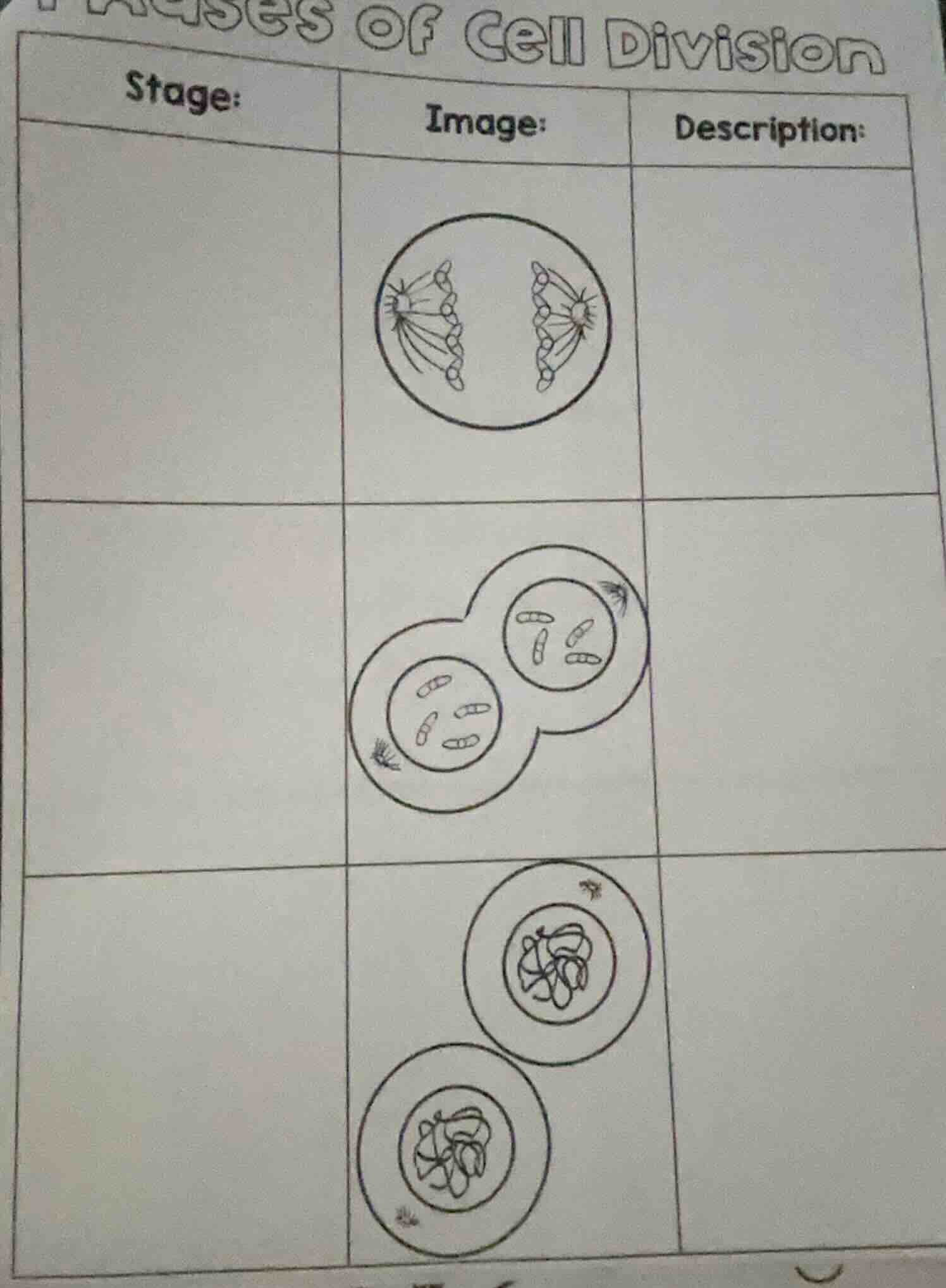
phases of cell division
stage:\timage:\tdescription:
\timage of a cell with chromosomes being pulled apart\t
\timage of two cells in the process of dividing, each with chromosomes\t
\timage of two cells with chromatin\t
To determine the stages of cell division (likely mitosis) for each image, we analyze the visual cues:
First Row (Top Image)
- Stage: Anaphase
- Description: Sister chromatids are pulled apart by spindle fibers toward opposite poles of the cell. The chromosomes are visibly separated and moving to each end of the cell.
Second Row (Middle Image)
- Stage: Cytokinesis (Telophase - late stage with division)
- Description: The cell has divided into two daughter cells, each with a nucleus containing the separated chromosomes. The cell membrane has pinched off (or a cell plate is forming, though this looks like animal cell cytokinesis with a cleavage furrow), resulting in two distinct cells.
Third Row (Bottom Image)
- Stage: Interphase (or Early Prophase, but more likely Interphase - specifically the stage with chromatin)
- Description: The cell(s) are in a resting/ growth phase. Chromatin is visible (uncondensed chromosomes), and the nucleus is intact. These cells are preparing for the next round of division or have completed division and are in interphase.
If you need to fill the table:
| Stage | Image Description |
|---|---|
| Cytokinesis | Two daughter cells formed, each with a nucleus and chromosomes; cell division done. |
| Interphase | Cells with intact nuclei, chromatin (uncondensed chromosomes) – resting/growth phase. |
(Note: The third row’s stage could also be early Prophase if chromatin is condensing, but Interphase is more common for the “resting” look with uncondensed genetic material.)

Snap & solve any problem in the app
Get step-by-step solutions on Sovi AI
Photo-based solutions with guided steps
Explore more problems and detailed explanations
To determine the stages of cell division (likely mitosis) for each image, we analyze the visual cues:
First Row (Top Image)
- Stage: Anaphase
- Description: Sister chromatids are pulled apart by spindle fibers toward opposite poles of the cell. The chromosomes are visibly separated and moving to each end of the cell.
Second Row (Middle Image)
- Stage: Cytokinesis (Telophase - late stage with division)
- Description: The cell has divided into two daughter cells, each with a nucleus containing the separated chromosomes. The cell membrane has pinched off (or a cell plate is forming, though this looks like animal cell cytokinesis with a cleavage furrow), resulting in two distinct cells.
Third Row (Bottom Image)
- Stage: Interphase (or Early Prophase, but more likely Interphase - specifically the stage with chromatin)
- Description: The cell(s) are in a resting/ growth phase. Chromatin is visible (uncondensed chromosomes), and the nucleus is intact. These cells are preparing for the next round of division or have completed division and are in interphase.
If you need to fill the table:
| Stage | Image Description |
|---|---|
| Cytokinesis | Two daughter cells formed, each with a nucleus and chromosomes; cell division done. |
| Interphase | Cells with intact nuclei, chromatin (uncondensed chromosomes) – resting/growth phase. |
(Note: The third row’s stage could also be early Prophase if chromatin is condensing, but Interphase is more common for the “resting” look with uncondensed genetic material.)