QUESTION IMAGE
Question
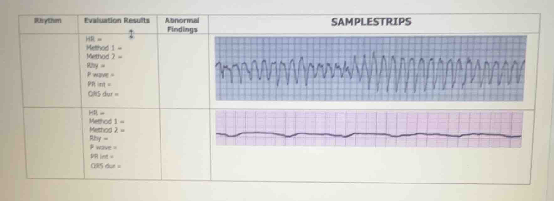
rhythm evaluation results abnormal findings samplestrips
hr =
method 1 =
method 2 =
rhy =
p wave =
pr int =
qrs dur =
hr =
method 1 =
method 2 =
rhy =
p wave =
pr int =
qrs dur =
Response
To analyze these ECG (electrocardiogram) strips, we'll evaluate each strip:
First (Top) ECG Strip:
- Rhythm: Appears chaotic, with no clear P waves, regular rhythm, or consistent QRS complexes. This is consistent with Ventricular Tachycardia (VT) or a more severe arrhythmia like Ventricular Fibrillation (VF) (though VF is usually more disorganized; this may be a rapid VT with aberrant conduction).
- HR (Heart Rate): Extremely fast (likely >150 bpm, possibly >200 bpm).
- Method 1/2 (Rate Calculation): Using the 300/number of large squares method: if we estimate, the R-R interval is very short (e.g., ~1 large square between peaks), so 300/1 = 300 bpm (unrealistic, so likely a calculation error due to irregularity—better to note the rhythm is irregular and fast).
- P wave: Absent (or indistinguishable due to rapid, chaotic activity).
- PR interval: Not measurable (no clear P waves or consistent PR segment).
- QRS duration: Wide (>0.12 seconds), consistent with ventricular origin.
- Abnormal Findings: Chaotic rhythm, wide QRS, absent P waves, extremely fast heart rate.
Second (Bottom) ECG Strip:
- Rhythm: Flat (isoelectric) line with no discernible P, QRS, or T waves. This is consistent with Asystole (no cardiac electrical activity) or a severely depressed rhythm (e.g., agonal rhythm, but agonal usually has small, irregular activity—this looks like straight asystole).
- HR: 0 (no detectable beats).
- Method 1/2: No R waves to count, so rate = 0.
- P wave, PR int, QRS dur: All unmeasurable (no electrical activity).
- Abnormal Findings: Complete absence of cardiac electrical activity (asystole).
Summary:
- Top strip: Likely Ventricular Tachycardia (VT) (or VF) with wide QRS, absent P waves, and rapid, chaotic rhythm.
- Bottom strip: Asystole (no electrical activity).
If you need to fill the table:
| Rhythm | Evaluation Results | Abnormal Findings | SAMPLESTRIPS (Visual) |
|---|---|---|---|
| Bottom: Asystole | HR = 0; Method 1/2 = N/A; Rhy = Absent; P wave = Absent; PR int = N/A; QRS dur = N/A | No cardiac electrical activity (asystole) | Flat line (isoelectric) |
Let me know if you need more specific interpretation (e.g., clinical context, next steps)!

Snap & solve any problem in the app
Get step-by-step solutions on Sovi AI
Photo-based solutions with guided steps
Explore more problems and detailed explanations
To analyze these ECG (electrocardiogram) strips, we'll evaluate each strip:
First (Top) ECG Strip:
- Rhythm: Appears chaotic, with no clear P waves, regular rhythm, or consistent QRS complexes. This is consistent with Ventricular Tachycardia (VT) or a more severe arrhythmia like Ventricular Fibrillation (VF) (though VF is usually more disorganized; this may be a rapid VT with aberrant conduction).
- HR (Heart Rate): Extremely fast (likely >150 bpm, possibly >200 bpm).
- Method 1/2 (Rate Calculation): Using the 300/number of large squares method: if we estimate, the R-R interval is very short (e.g., ~1 large square between peaks), so 300/1 = 300 bpm (unrealistic, so likely a calculation error due to irregularity—better to note the rhythm is irregular and fast).
- P wave: Absent (or indistinguishable due to rapid, chaotic activity).
- PR interval: Not measurable (no clear P waves or consistent PR segment).
- QRS duration: Wide (>0.12 seconds), consistent with ventricular origin.
- Abnormal Findings: Chaotic rhythm, wide QRS, absent P waves, extremely fast heart rate.
Second (Bottom) ECG Strip:
- Rhythm: Flat (isoelectric) line with no discernible P, QRS, or T waves. This is consistent with Asystole (no cardiac electrical activity) or a severely depressed rhythm (e.g., agonal rhythm, but agonal usually has small, irregular activity—this looks like straight asystole).
- HR: 0 (no detectable beats).
- Method 1/2: No R waves to count, so rate = 0.
- P wave, PR int, QRS dur: All unmeasurable (no electrical activity).
- Abnormal Findings: Complete absence of cardiac electrical activity (asystole).
Summary:
- Top strip: Likely Ventricular Tachycardia (VT) (or VF) with wide QRS, absent P waves, and rapid, chaotic rhythm.
- Bottom strip: Asystole (no electrical activity).
If you need to fill the table:
| Rhythm | Evaluation Results | Abnormal Findings | SAMPLESTRIPS (Visual) |
|---|---|---|---|
| Bottom: Asystole | HR = 0; Method 1/2 = N/A; Rhy = Absent; P wave = Absent; PR int = N/A; QRS dur = N/A | No cardiac electrical activity (asystole) | Flat line (isoelectric) |
Let me know if you need more specific interpretation (e.g., clinical context, next steps)!