QUESTION IMAGE
Question
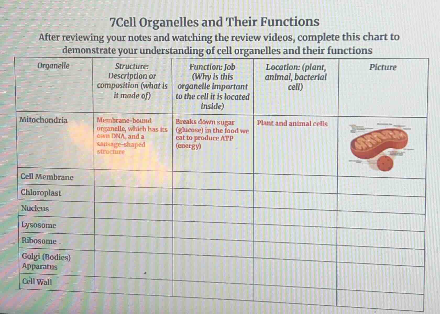
7cell organelles and their functions
after reviewing your notes and watching the review videos, complete this chart to demonstrate your understanding of cell organelles and their functions
| organelle | structure: description or composition (what is it made of) | function: job (why is this organelle important to the cell it is located inside) | location: (plant, animal, bacterial cell) | picture |
|---|---|---|---|---|
| cell membrane | ||||
| chloroplast | ||||
| nucleus | ||||
| lysosome | ||||
| ribosome | ||||
| golgi (bodies) apparatus | ||||
| cell wall |
To complete the chart for each cell organelle, we use knowledge of cell biology (subfield of Biology, Natural Science) to fill in the structure, function, location, and (if applicable) picture description. Here's the breakdown for each organelle:
1. Cell Membrane
- Structure: A thin, flexible, semi - permeable lipid bilayer (made of phospholipids, proteins, cholesterol, and carbohydrates) with a fluid - mosaic structure.
- Function: Regulates the entry and exit of substances (like nutrients, waste, and ions) into and out of the cell; also provides structural support and cell recognition.
- Location: Plant, animal, and bacterial cells (all cells have a cell membrane).
- Picture: Typically depicted as a double - layered line (or a more detailed fluid - mosaic model with embedded proteins) surrounding the cell’s interior.
2. Chloroplast
- Structure: Membrane - bound organelle with an outer membrane, inner membrane, and a system of thylakoids (stacked into grana) in a stroma - filled matrix; contains chlorophyll (pigment) and its own DNA.
- Function: Captures light energy from the sun and uses it to convert carbon dioxide and water into glucose (sugar) and oxygen through photosynthesis.
- Location: Plant cells (and some photosynthetic bacteria, but mainly plant cells).
- Picture: Usually shown as a green, oval - shaped organelle with internal stacks (grana) of thylakoids.
3. Nucleus
- Structure: Membrane - bound (nuclear envelope with pores) organelle containing the cell’s DNA (in the form of chromatin or chromosomes) and a nucleolus (where ribosomes are made).
- Function: Controls the cell’s activities (growth, metabolism, reproduction) by regulating gene expression; stores genetic information.
- Location: Plant and animal cells (eukaryotic cells; bacterial cells lack a nucleus).
- Picture: A circular or oval - shaped structure with a darker - stained nucleolus inside, surrounded by a nuclear envelope.
4. Lysosome
- Structure: Membrane - bound vesicle containing digestive enzymes (hydrolytic enzymes).
- Function: Digests (breaks down) waste materials, old cell parts (organelles), and foreign invaders (like bacteria) in the cell; helps in cell renewal and defense.
- Location: Animal cells (rare in plant cells, where vacuoles perform similar functions).
- Picture: Depicted as a small, spherical vesicle with enzymes inside, often near the Golgi apparatus.
5. Ribosome
- Structure: Small, non - membrane - bound organelle made of ribosomal RNA (rRNA) and proteins; exists as free ribosomes (in the cytoplasm) or bound ribosomes (attached to the rough endoplasmic reticulum).
- Function: Synthesizes proteins by translating messenger RNA (mRNA) into polypeptide chains (amino acid sequences).
- Location: Plant, animal, and bacterial cells (all cells need protein synthesis).
- Picture: Shown as tiny dots (or small spheres) in the cytoplasm or attached to the rough ER.
6. Golgi (Bodies) Apparatus
- Structure: A stack of flattened, membrane - bound sacs (cisternae) with associated vesicles.
- Function: Modifies, sorts, and packages proteins (from the rough ER) and lipids; transports these molecules to their final destinations (inside the cell, outside the cell, or to other organelles).
- Location: Plant and animal cells (eukaryotic cells).
- Picture: Drawn as a stack of pancakes - like sacs with vesicles budding off.
7. Cell Wall
- Structure: A rigid, outer layer made of cellulose (in plant…

Snap & solve any problem in the app
Get step-by-step solutions on Sovi AI
Photo-based solutions with guided steps
Explore more problems and detailed explanations
To complete the chart for each cell organelle, we use knowledge of cell biology (subfield of Biology, Natural Science) to fill in the structure, function, location, and (if applicable) picture description. Here's the breakdown for each organelle:
1. Cell Membrane
- Structure: A thin, flexible, semi - permeable lipid bilayer (made of phospholipids, proteins, cholesterol, and carbohydrates) with a fluid - mosaic structure.
- Function: Regulates the entry and exit of substances (like nutrients, waste, and ions) into and out of the cell; also provides structural support and cell recognition.
- Location: Plant, animal, and bacterial cells (all cells have a cell membrane).
- Picture: Typically depicted as a double - layered line (or a more detailed fluid - mosaic model with embedded proteins) surrounding the cell’s interior.
2. Chloroplast
- Structure: Membrane - bound organelle with an outer membrane, inner membrane, and a system of thylakoids (stacked into grana) in a stroma - filled matrix; contains chlorophyll (pigment) and its own DNA.
- Function: Captures light energy from the sun and uses it to convert carbon dioxide and water into glucose (sugar) and oxygen through photosynthesis.
- Location: Plant cells (and some photosynthetic bacteria, but mainly plant cells).
- Picture: Usually shown as a green, oval - shaped organelle with internal stacks (grana) of thylakoids.
3. Nucleus
- Structure: Membrane - bound (nuclear envelope with pores) organelle containing the cell’s DNA (in the form of chromatin or chromosomes) and a nucleolus (where ribosomes are made).
- Function: Controls the cell’s activities (growth, metabolism, reproduction) by regulating gene expression; stores genetic information.
- Location: Plant and animal cells (eukaryotic cells; bacterial cells lack a nucleus).
- Picture: A circular or oval - shaped structure with a darker - stained nucleolus inside, surrounded by a nuclear envelope.
4. Lysosome
- Structure: Membrane - bound vesicle containing digestive enzymes (hydrolytic enzymes).
- Function: Digests (breaks down) waste materials, old cell parts (organelles), and foreign invaders (like bacteria) in the cell; helps in cell renewal and defense.
- Location: Animal cells (rare in plant cells, where vacuoles perform similar functions).
- Picture: Depicted as a small, spherical vesicle with enzymes inside, often near the Golgi apparatus.
5. Ribosome
- Structure: Small, non - membrane - bound organelle made of ribosomal RNA (rRNA) and proteins; exists as free ribosomes (in the cytoplasm) or bound ribosomes (attached to the rough endoplasmic reticulum).
- Function: Synthesizes proteins by translating messenger RNA (mRNA) into polypeptide chains (amino acid sequences).
- Location: Plant, animal, and bacterial cells (all cells need protein synthesis).
- Picture: Shown as tiny dots (or small spheres) in the cytoplasm or attached to the rough ER.
6. Golgi (Bodies) Apparatus
- Structure: A stack of flattened, membrane - bound sacs (cisternae) with associated vesicles.
- Function: Modifies, sorts, and packages proteins (from the rough ER) and lipids; transports these molecules to their final destinations (inside the cell, outside the cell, or to other organelles).
- Location: Plant and animal cells (eukaryotic cells).
- Picture: Drawn as a stack of pancakes - like sacs with vesicles budding off.
7. Cell Wall
- Structure: A rigid, outer layer made of cellulose (in plant cells), peptidoglycan (in bacterial cells), or chitin (in fungal cells); provides structural support.
- Function: Gives the cell shape, protects the cell from mechanical damage and osmotic stress; prevents the cell from bursting in a hypotonic environment.
- Location: Plant cells, bacterial cells, and fungal cells (animal cells lack a cell wall).
- Picture: Depicted as a thick, outer layer surrounding the cell membrane in plant/bacterial cells.
To fill the chart, you can transfer this information into the respective columns. For example:
| Organelle | Structure | Function | Location | Picture (Description) |
|---|---|---|---|---|
| Chloroplast | Membrane - bound, thylakoids (grana), stroma, chlorophyll, own DNA | Photosynthesis (makes glucose + O₂) | Plant cells | Green, oval with internal grana |
| Nucleus | Membrane - bound, chromatin/DNA, nucleolus, nuclear pores | Controls cell activities; stores DNA | Plant, animal cells | Circular with dark nucleolus, nuclear envelope |
| Lysosome | Membrane - bound vesicle, digestive enzymes | Digests waste, old parts, invaders | Animal cells | Small vesicle with enzymes |
| Ribosome | Non - membrane - bound, rRNA + proteins, free or bound | Protein synthesis (translates mRNA) | Plant, animal, bacterial cells | Tiny dots (free) or attached to rough ER |
| Golgi (Bodies) Apparatus | Stack of flattened cisternae, vesicles | Modifies, sorts, packages proteins/lipids | Plant, animal cells | Stack of “pancakes” with budding vesicles |
| Cell Wall | Rigid, cellulose (plant), peptidoglycan (bacteria) | Supports, protects, prevents bursting | Plant, bacterial, fungal cells | Thick outer layer around cell membrane |
If you need to draw the pictures, use the descriptions to guide your illustrations (e.g., chloroplast as green ovals with grana, mitochondria as sausage - shaped with inner folds, etc.).