QUESTION IMAGE
Question
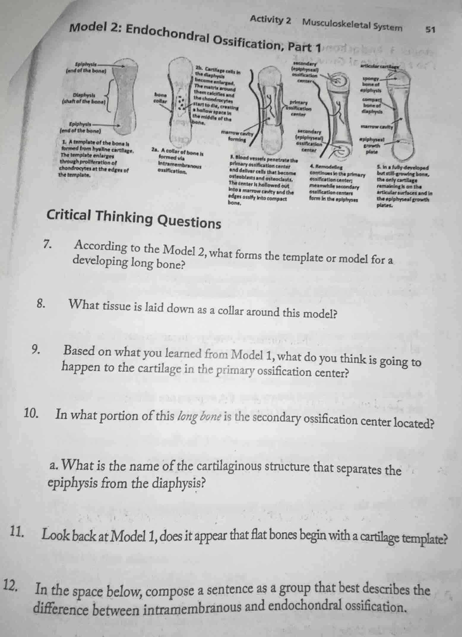
model 2: endochondral ossification, part 1
activity 2 musculoskeletal system
51
(diaphysis (shaft of the bone)
epiphysis (end of the bone)
epiphysis (end of the bone)
- a template of the bone is formed from hyaline cartilage. the template enlarges through proliferation of chondrocytes at the edges of the template.
bone collar
2a. a collar of bone is formed via intramembranous ossification.
2b. cartilage cells in the diaphysis become enlarged. the matrix around them calcifies and the chondrocytes start to die, creating a hollow space in the middle of the bone.
primary ossification center
marrow cavity forming
- blood vessels penetrate the primary ossification center and deliver cells that become osteoblasts and osteoclasts. the center is hollowed out into a marrow cavity and the edges ossify into compact bone.
secondary (epiphyseal) ossification center
secondary (epiphyseal) ossification center
- remodeling continues in the primary ossification center; meanwhile secondary ossification centers form in the epiphyses
articular cartilage
spongy bone of epiphysis
compact bone of diaphysis
marrow cavity
s. in a fully-developed but still-growing bone, the only cartilage remaining is on the articular surfaces and in the epiphyseal growth plates.
epiphyseal growth plate
critical thinking questions
- according to the model 2, what forms the template or model for a developing long bone?
- what tissue is laid down as a collar around this model?
- based on what you learned from model 1, what do you think is going to happen to the cartilage in the primary ossification center?
- in what portion of this long bone is the secondary ossification center located?
a. what is the name of the cartilaginous structure that separates the epiphysis from the diaphysis?
- look back at model 1, does it appear that flat bones begin with a cartilage template?
- in the space below, compose a sentence as a group that best describes the difference between intramembranous and endochondral ossification.
Question 7
From Model 2 (step 1), it says a template of the bone is formed from hyaline cartilage. So we refer to that description.
In Model 2 (step 2a), it states a collar of bone is formed via intramembranous ossification around the model. So the tissue laid down as a collar is bone (formed through intramembranous ossification, so the tissue is bone tissue).
In Model 2 (step 2b), cartilage cells in the diaphysis become enlarged, the matrix around them calcifies, and the chondrocytes start to die, creating a hollow space. Also, from the process of endochondral ossification, in the primary ossification center, cartilage is replaced by bone as osteoblasts and osteoclasts act. So the cartilage in the primary ossification center will be replaced by bone (or calcify, die, and be replaced as part of the ossification process).

Snap & solve any problem in the app
Get step-by-step solutions on Sovi AI
Photo-based solutions with guided steps
Explore more problems and detailed explanations
Hyaline cartilage forms the template or model for a developing long bone.